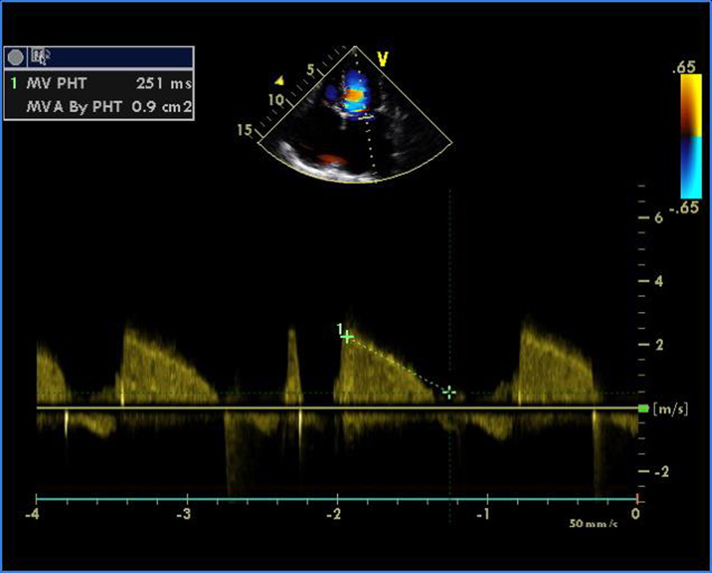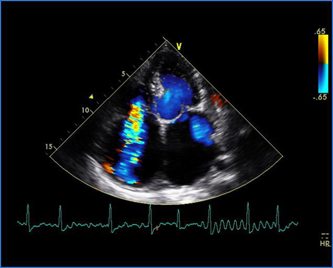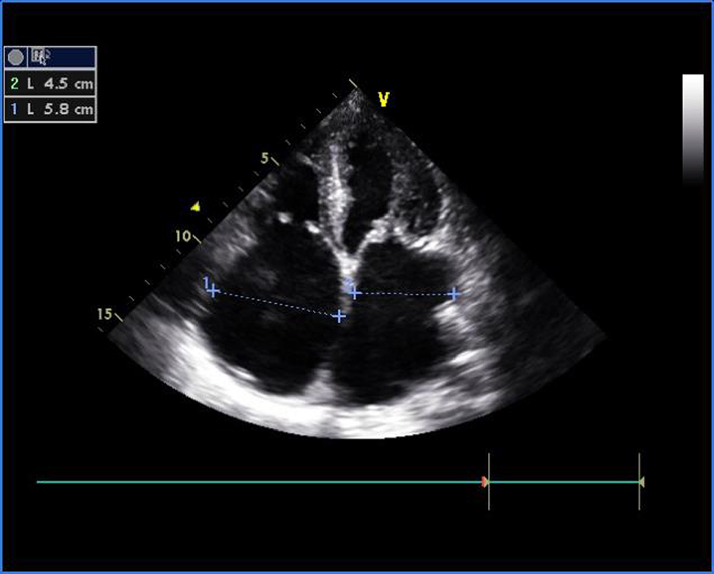
Figure 1. Transthoracic echocardiogram of apical four-chamber view showing thickened and doming mitral valve and tricuspid valve.
| Cardiology Research, ISSN 1923-2829 print, 1923-2837 online, Open Access |
| Article copyright, the authors; Journal compilation copyright, Cardiol Res and Elmer Press Inc |
| Journal website https://www.cardiologyres.org |
Case Report
Volume 6, Number 6, December 2015, pages 357-361
Malignant Rheumatic Heart Disease Presenting as Quadrivalvular Stenosis
Figures