Cardiology Research, ISSN 1923-2829 print, 1923-2837 online, Open Access
Article copyright, the authors; Journal compilation copyright, Cardiol Res and Elmer Press Inc
Journal website http://www.cardiologyres.org

Case Report

Volume 11, Number 2, April 2020, pages 129-133


Primary Cardiac Undifferentiated High-Grade Intimal Pleomorphic Sarcoma: A Case Series Report

Figures

Figure 1.
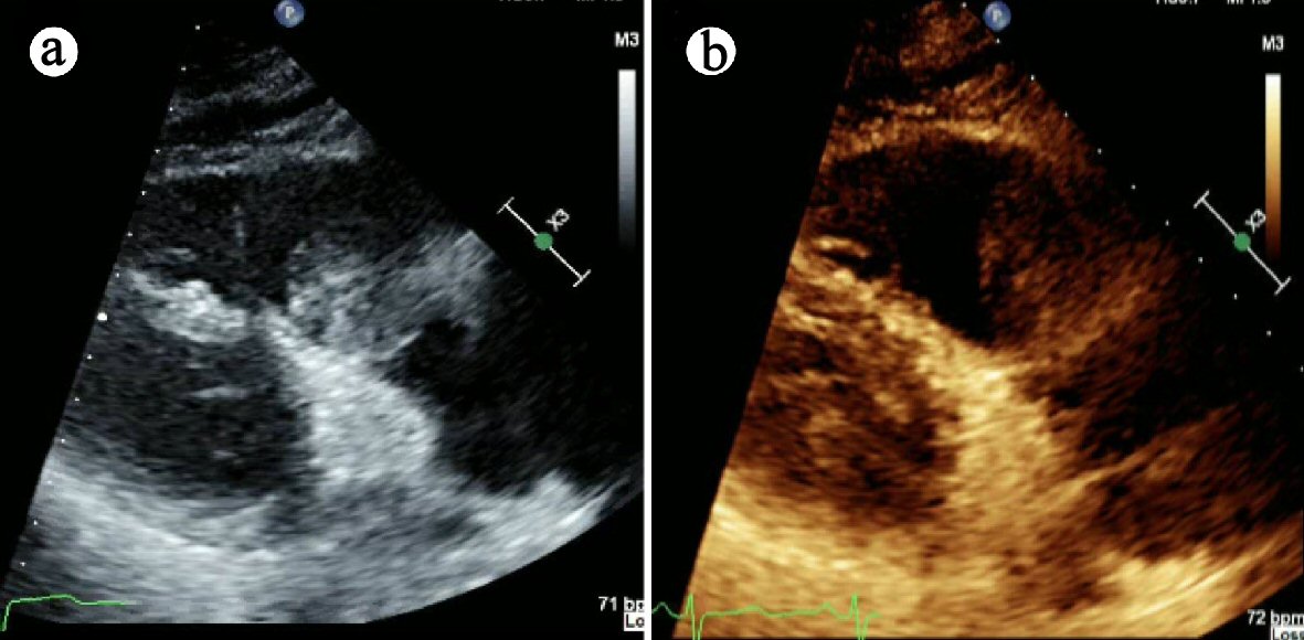
Figure 1. Case 1: TTE demonstrating large mobile, well circumscribed echo-density in the right ventricular outflow tract (RVOT) measuring 6 × 2 cm. TTE: transthoracic echocardiogram.
Figure 2.
Figure 2. (a) Case 1: CT angiogram demonstrating large intraluminal mass centered in the RVOT. (b) Large eccentric mural thrombus in right main pulmonary artery with smaller filling defects in the distal segmental branches. CT: computed tomography; RVOT: right ventricular outflow tract.
Figure 3.
Figure 3. (a) Case 2: TTE demonstrating mass attached to the intraatrial septum adjacent to anterior mitral annulus. (b) Atrial tumor cross section. (c) Pleomorphic, spindle-shaped cells with hyper-chromatic nuclei and prominent mitotic figures. TTE: transthoracic echocardiogram.
Figure 4.
Figure 4. (a) Case 2: TEE demonstrating recurrence of mobile heterogenous echodense mass in left atrium at 20-month interval. (b) Hypermetabolic left atrial lesion on PET/CT. (c) Mass adjacent to anterior mitral leaflet (and prolapses into left ventricle) on CMR. (d) Mass within the left atrium on CMR. TEE: transesophageal echocardiogram; PET/CT: positron emission tomography/computed tomography; CMR: cardiovascular magnetic resonance imaging.