Cardiology Research, ISSN 1923-2829 print, 1923-2837 online, Open Access
Article copyright, the authors; Journal compilation copyright, Cardiol Res and Elmer Press Inc
Journal website https://www.cardiologyres.org

Original Article

Volume 13, Number 5, October 2022, pages 289-296


Carotid Artery Revascularization Improves Cardiac Sympathetic Nerve Activity in Patients With Carotid Artery Stenosis

Figures

Figure 1.
Figure 1. Patients and protocol. ECG: electrocardiography; 123I-MIBG scintigraphy: iodine-123-metaiodobezylguanidine scintigraphy; ABPM: ambulatory blood pressure monitoring; TTE: transthoracic echocardiography; CEA: carotid endarterectomy; CAS: carotid artery stenting.
Figure 2.
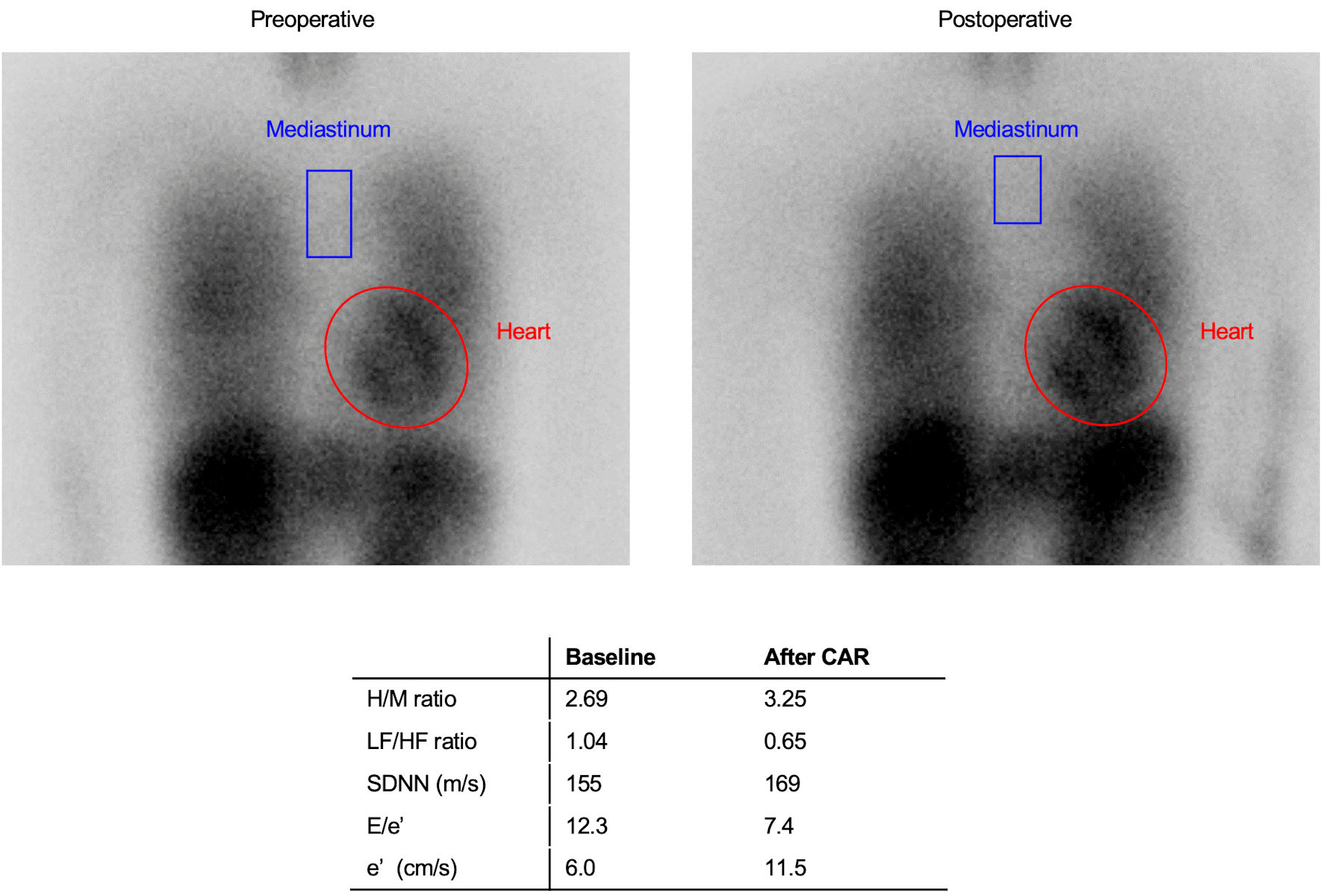
Figure 2. Representative case of improvement in sympathetic function after CAR. A 57-year-old woman with general cardiovascular risk factors, including hypertension and dyslipidemia was not taking beta-blockers. The H/M ratio on 123I-MIBG scintigraphy was calculated by dividing the mean pixel value within the myocardium by the mean pixel value within the mediastinum, using the region of interest (ROI) of the entire heart and the upper mediastinum on the planar image. Compared to pre-CAR, MIBG uptake in the heart was higher than in the upper mediastinum, suggesting decreased sympathetic nerve activity. CAR: carotid artery revascularization; H/M: heart-to-mediastinum; 123I-MIBG: iodine-123-metaiodobezylguanidine; LF: low-frequency; HF: high-frequency; SDNN: standard deviation of the N-N interval.
Figure 3.
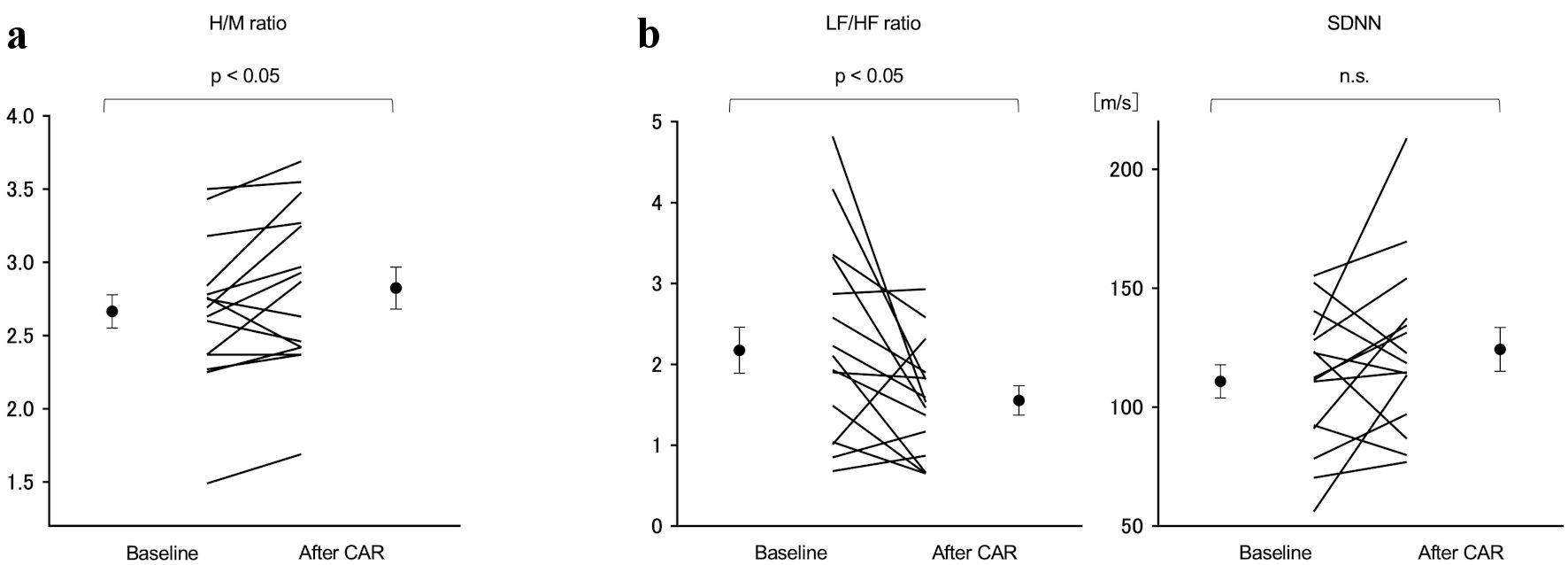
Figure 3. Comparison of the H/M ratio on the 123I MIBG scintigraphy and LF/HF ratio and SDNN on 24-h Holter ECG pre- and post-CAR. (a) The H/M ratio increased significantly from 2.66 ± 0.48 preoperatively to 2.86 ± 0.56 postoperatively (P < 0.05). (b) The LF/HF ratio decreased significantly from 2.17 ± 1.20 preoperatively to 1.62 ± 0.68 postoperatively (P < 0.04). SDNN showed no change. H/M: heart-to-mediastinum; 123I-MIBG: iodine-123-metaiodobezylguanidine; LF: low-frequency; HF: high-frequency; SDNN: standard deviation of the N-N interval; CAR: carotid artery revascularization.
Figure 4.
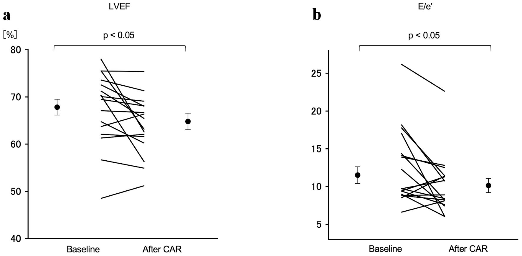
Figure 4. Comparison of LVEF and E/e’ between pre- and post-CAR. (a) The LVEF decreased significantly from 67.8±7.7% preoperatively to 64.8±7.2% postoperatively (P < 0.05). (b) The E/e’ decreased significantly from 11.7 ± 5.1 preoperatively to 10.1 ± 4.0 postoperatively (P < 0.05). LVEF: left ventricular ejection fraction; CAR: carotid artery revascularization.

Tables

Table 1. Patient Characteristics
 
N = 21
Hb: hemoglobin; Cre: creatinine; TG: triglyceride; LDL: low-density lipoprotein; HDL: high-density lipoprotein; HbA1c: hemoglobin A1c; CCB: calcium channel blocker; ACE-I: angiotensin-converting enzyme inhibitor; ARB: angiotensin receptor blocker.
Age (year)69.5 ± 6.8
Male gender, n (%)18 (86)
Body mass index (kg/m2)22.5 ± 4.0
Hypertension, n (%)17 (81)
Diabetes, n (%)7 (33)
Dyslipidemia, n (%)20 (95)
Smoking, n (%)5 (24)
Renal failure, n (%)7 (33)
Sleep apnea syndrome, n (%)0 (0)
Laboratory data
  Hb (g/dL)12.3 (11.6 - 13.9)
  Cre (mg/dL)0.9 (0.8 - 1.1)
  TG (mg/dL)106 (64.5 - 139.5)
  HDL (mg/dL)53 (42 - 78)
  LDL (mg/dL)66 (48 - 88)
  HbA1c (%)5.6 (5.4 - 6.2)
Medication
  CCB9 (43)
  ACE-I/ARB9 (43)
  β-blocker3 (14)
  Diuretics1 (5)


Table 2. Comparison of Common Carotid Artery Flow Before and After Revascularization
 
BaselineAfter CARP value
CAR: carotid artery revascularization; CCA: common carotid artery; EDV: end-diastolic velocity.
CCA-EDV (cm/s)10.8 ± 3.216.1 ± 7.1< 0.01
CCA-EDV ratio1.68 ± 0.541.2 ± 0.14< 0.01


Table 3. Comparison of Pre- and Postoperative Echocardiographic Data and Laboratory Data
 
BaselineAfter CARP value
CAR: carotid artery revascularization; LVEF: left ventricular ejection fraction; LVDd: left ventricular diastolic diameter; LVMI: left ventricular mass index; LAD: left atrial diameter.
Echocardiography
  LVEF (%)67.8 ± 7.764.8 ± 7.2< 0.05
  LVDd (mm)46.5 ± 4.745.2 ±4.30.6
  LVMI128 ± 62127 ± 540.38
  LAD (mm)40.0 ± 6.539.5 ± 6.40.76
  E/e’11.5 ± 5.110.1 ± 3.9< 0.05
  e’ (cm/s)5.9 ± 1.96.3 ± 1.80.17
Laboratory data
  BNP (pg/mL)34.2 ± 26.430.2 ± 25.70.48


Table 4. Comparison of Pre- and Postoperative ABPM Data
 
BaselineAfter CARP value
aExtreme-dipper: with ≥ 20% nocturnal systolic blood pressure fall. bDipper: with ≥ 10% but < 20% fall. cNon-dipper: with ≥ 0% but < 10% fall. dRiser: with < 0% fall. ABPM: ambulatory blood pressure monitoring; CAR: carotid artery revascularization; BP: blood pressure.
Extreme-dippera, n (%)2 (13)2 (13)0.2
Dipperb, n (%)7 (47)6 (40)
Non-dipperc, n (%)3 (20)7 (47)
Riserd, n (%)3 (20)0 (0)
24-h BP (systole), mm Hg126 ± 21133 ± 120.27
24-h BP (diastole), mm Hg70 ± 1274 ± 80.17
Night-time BP (systole), mm Hg122 ± 23123 ± 210.22
Night-time BP (diastole), mm Hg67 ± 1369 ± 100.71