Figures
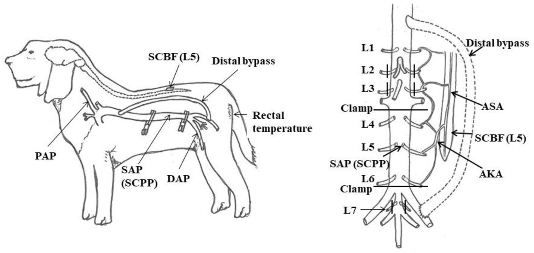
Figure 1. Experimental model. AKA: artery of Adamkiewicz; ASA: anterior spinal artery; DAP: distal arterial blood pressure; PAP: proximal arterial blood pressure; SAP: segmental arterial pressure; SCBF: spinal cord blood flow; SCPP: spinal cord perfusion pressure. Reproduced from Journal of Thoracic and Cardiovascular Surgery, in press [9]. ©Elsevier.
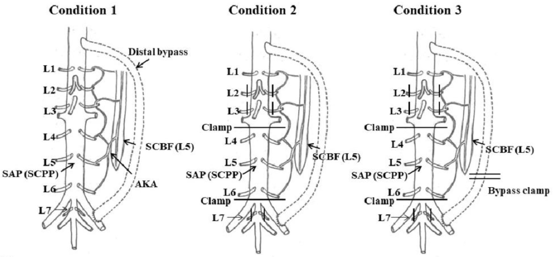
Figure 2. Experimental protocol. Condition 1: no aortic clamp and no SA clamp (control group). Condition 2: L2-L7 SA flow halted with distal perfusion. Condition 3: L2-L7 SA flow halted with no distal perfusion. AKA: artery of Adamkiewicz; SAP: segmental arterial pressure; SCBF: spinal cord blood flow; SCPP: spinal cord perfusion pressure. Reproduced from Journal of Thoracic and Cardiovascular Surgery, in press [9]. ©Elsevier.
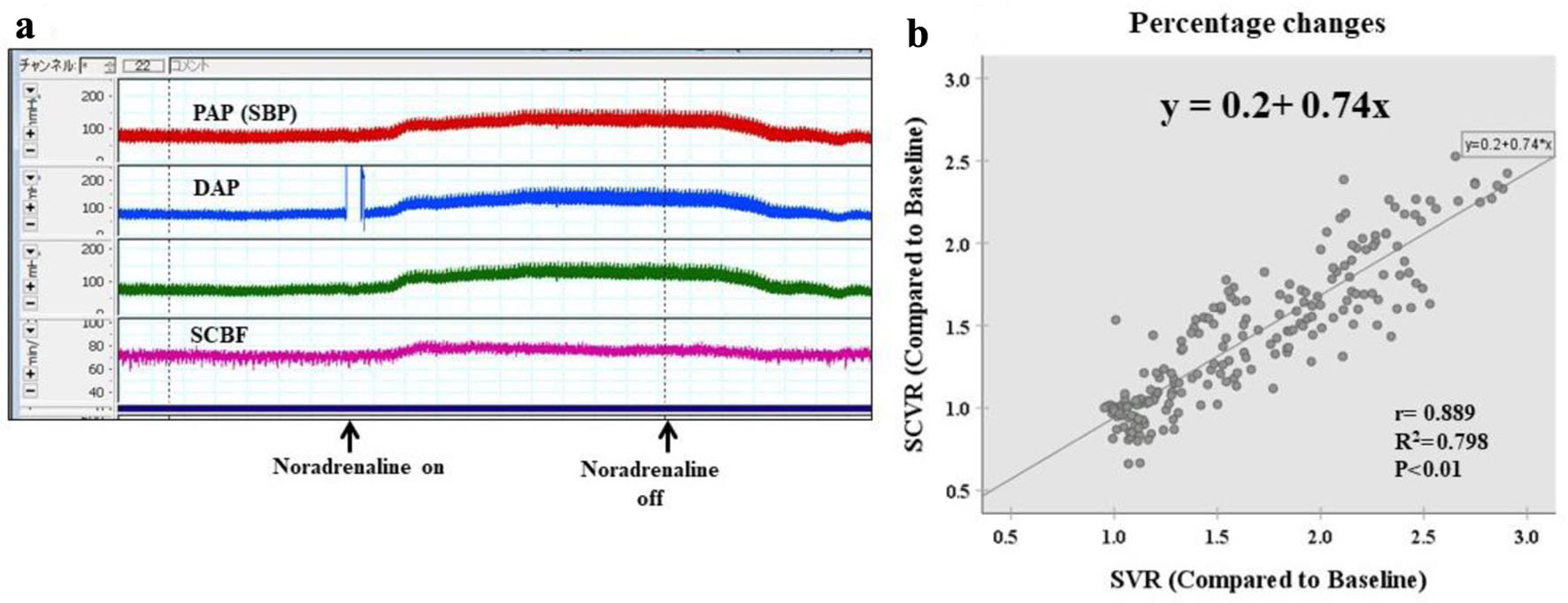
Figure 3. Condition 1: no aortic clamp and no SA clamp (no spinal ischemia: control group). (a) Laboratory chart shows the real-time record of SBP, DAP, and SCBF. SCBF increased in accordance with the increase in SBP induced by noradrenaline administration. (b) Scattergram of percentage changes shows a strong positive correlation between SVR and SCVR, and simple regression analysis yields an equation that relates the two variables with high predictive accuracy, with a coefficient of determination of 0.79 (y = 0.2 + 0.74x, r = 0.889, r2 = 0.789, P < 0.01). DAP: distal aortic blood pressure; PAP: proximal arterial blood pressure; SBP: systemic blood pressure; SCBF: spinal cord blood flow; SCVR: spinal cord vascular resistance; SVR: systematic vascular resistance.
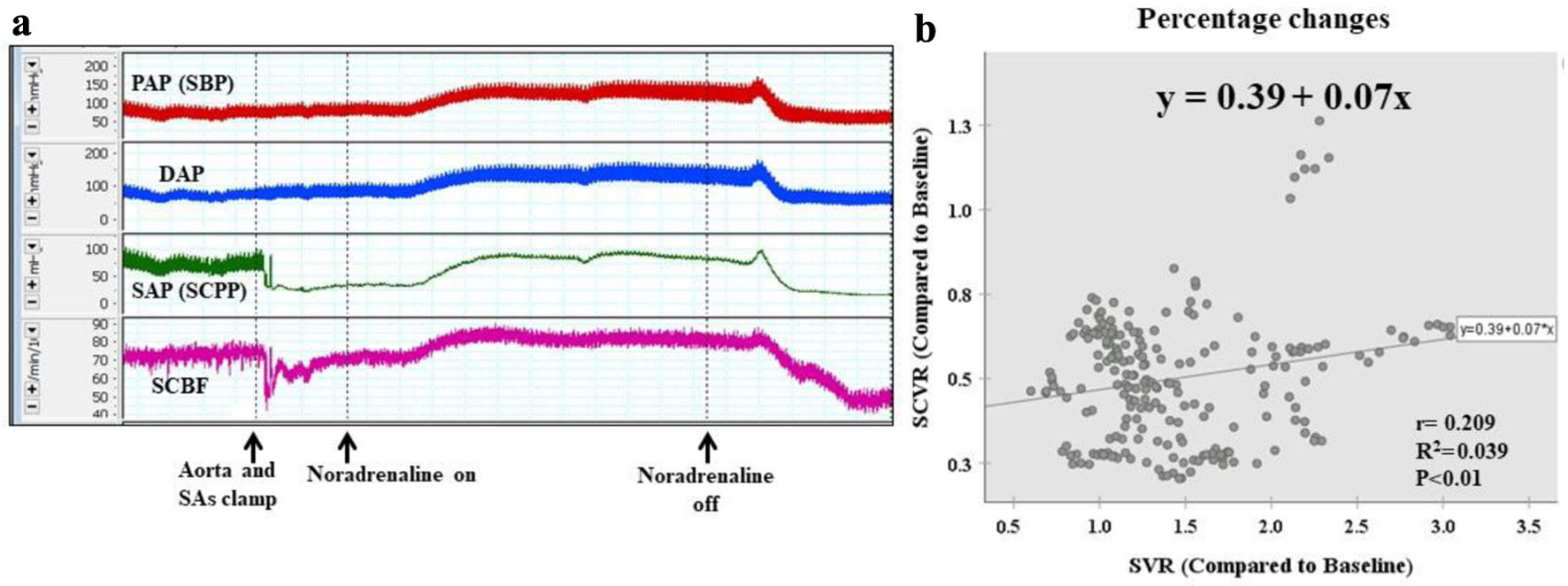
Figure 4. Condition 2: six pairs of SAs clamped (with distal perfusion). (a) When inflow from the SAs is stopped, SCBF and SAP (SCPP) decrease. However, they increase as systemic blood pressure increases. (b) Scattergram of percentage changes shows a weak correlation between the rate of change in SVR and the rate of change in SCVR (y = 0.39 + 0.07x, r = 0.209, r2 = 0.039, P < 0.01). The rate of increase in SCVR is far lower than the rate of increase in SVR. DAP: distal arterial blood pressure; PAP: proximal arterial blood pressure; SA: segmental artery; SAP: segmental arterial pressure; SBP: systemic blood pressure; SCBF: spinal cord blood flow; SCPP: spinal cord perfusion pressure; SCVR: spinal cord vascular resistance; SVR: systematic vascular resistance.
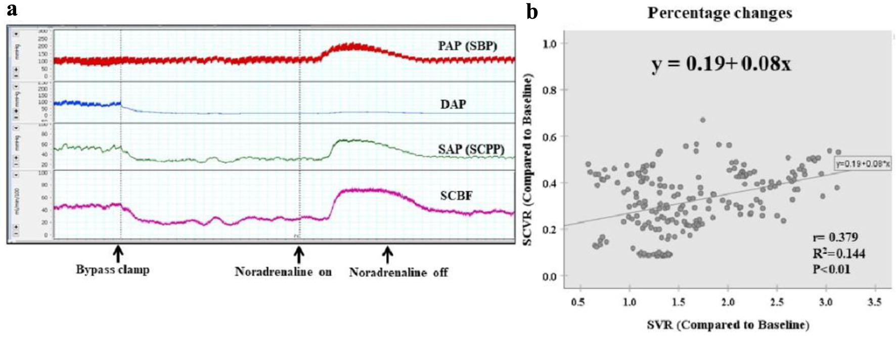
Figure 5. Condition 3: six pairs of SAs clamped (without distal perfusion). (a) When the distal bypass is clamped, SCBF and SAP (SCPP) decrease further. However, they then increase as systemic blood pressure increases. (b) Scattergram of percentage changes shows a weak correlation between the rate of change in SVR and the rate of change in SCVR (y = 0.19 + 0.08x, r = 0.379, r2 = 0.144, P < 0.01). Without distal perfusion, the rate of increase in SCVR is even lower than under condition 2. DAP: distal arterial blood pressure; PAP: proximal arterial blood pressure; SAs: segmental arteries; SAP: segmental arterial pressure; SBP: systemic blood pressure; SCBF: spinal cord blood flow; SCPP: spinal cord perfusion pressure; SCVR: spinal cord vascular resistance; SVR: systematic vascular resistance.