Cardiology Research, ISSN 1923-2829 print, 1923-2837 online, Open Access
Article copyright, the authors; Journal compilation copyright, Cardiol Res and Elmer Press Inc
Journal website https://www.cardiologyres.org

Case Report

Volume 14, Number 2, April 2023, pages 153-157


Left Axillary Access for Transcatheter Aortic Valve Implantation in a Patient With Two Dependent Internal Mammary Artery Grafts and a Permanent Left-Sided Implanted Pacemaker

Figures

Figure 1.
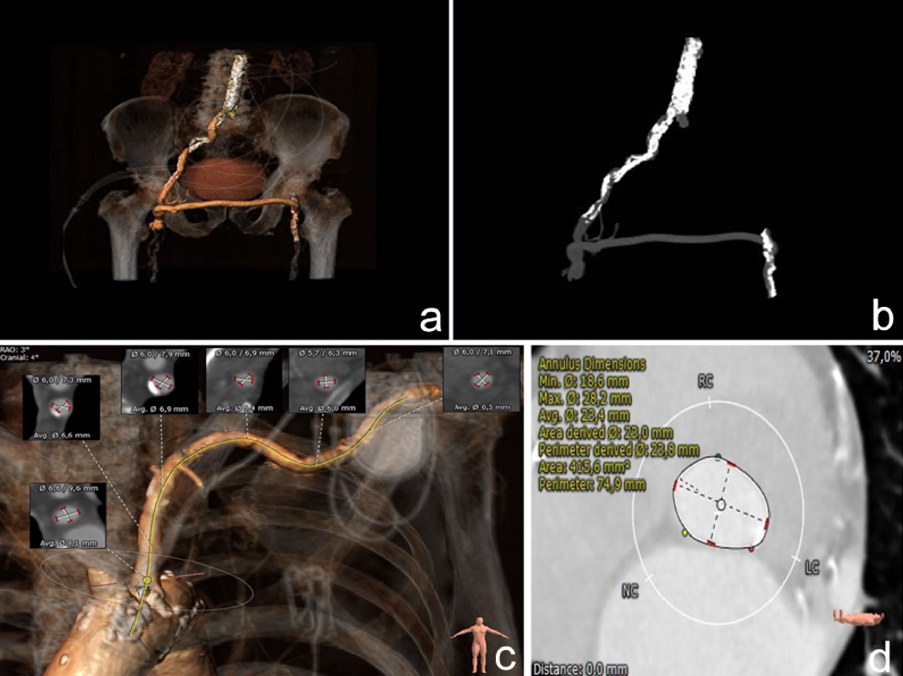
Figure 1. (a) Three-dimensional (3D) reconstruction of the aortoiliofemoral anatomy and (b) intravenous (IV) contrast image from MSCT scanning. Severe bilateral femoral artery disease with a patent femoro-femoral bypass graft and severe calcifications of the right aortoiliac artery and the descending aorta are displayed. (c) 3D reconstruction of the left subclavian artery anatomy and regional vessel diameters, with permanent pacemaker implanted beside the left acromioclavicular joint. (d) Aortic annulus perimeter from MSCT scanning. MSCT: multislice computed tomography.
Figure 2.
Figure 2. (a) Successful safe cutdown approach of the left axillary artery sparing the surrounding anatomical structures and the pacemaker/lead pocket and (b) subsequent insertion of a 14-F vascular sheath.
Figure 3.
Figure 3. (a) Fluoroscopy injection confirming the non-occlusive position of the sheath and patency of the LIMA graft. (b) A 27-mm self-expandable transcatheter aortic valve stationed over a stiff guide wire at the level of the aortic annulus. (c) Fully expanded valve with (d) no significant angiographic tracings of aortic regurgitation and/or paravalvular leak. LIMA: left internal mammary artery.