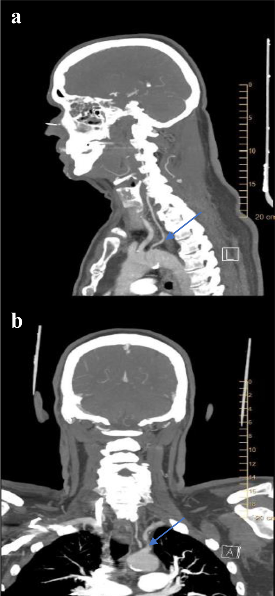
Figure 2. CT angiogram of neck showing (a) the anomalous origin of the left vertebral artery directly from the aortic arch between the left common carotid artery and the left subclavian artery (LSCA) (blue arrow), and (b) proximal LSCA stenosis (blue arrow).
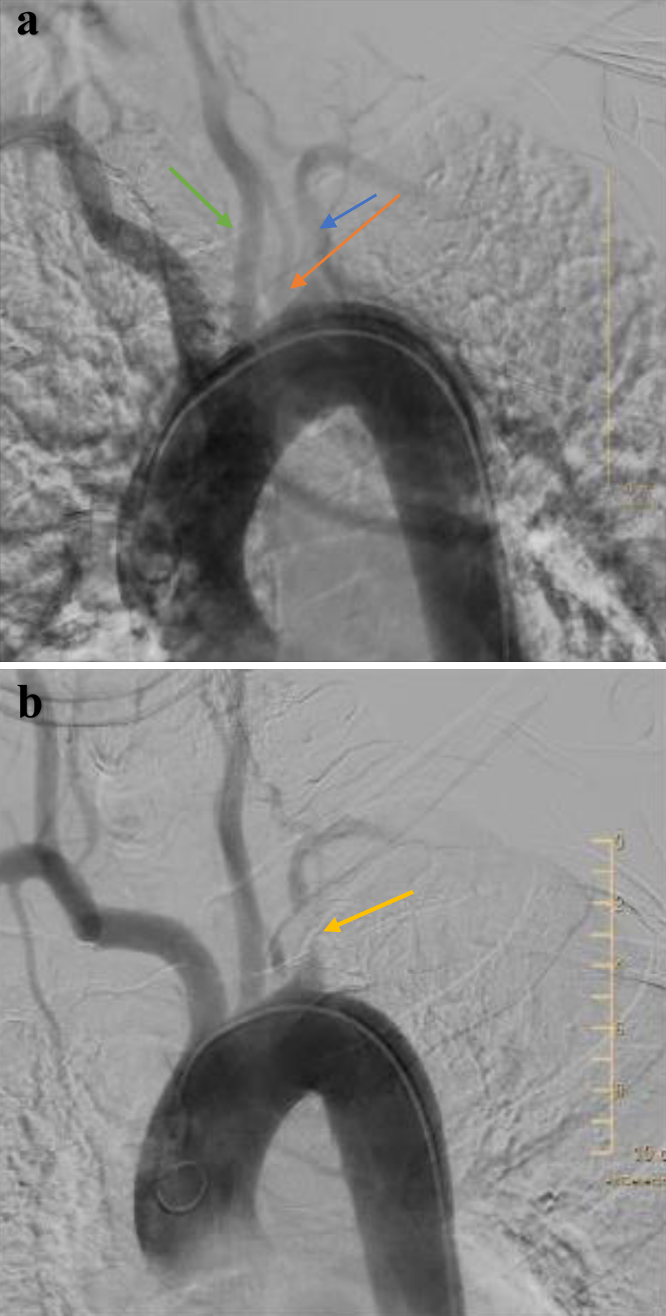
Figure 3. Aortic arch angiogram showing (a) the anomalous origin of the left vertebral artery (orange arrow) directly from the aortic arch between the left common carotid artery (green arrow) and the left subclavian artery (LSCA) (blue arrow), and (b) proximal LSCA stenosis.
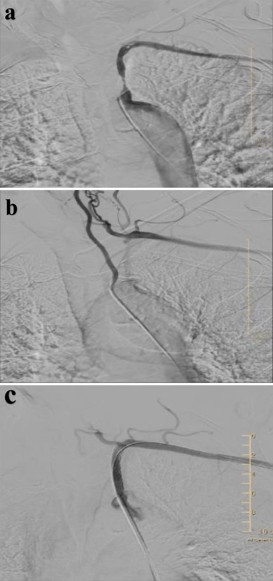
Figure 4. (a) Selective angiography confirming significant stenosis of the proximal left subclavian artery (LSCA) with no flow in thyrocervical trunk, (b) selective angiography of left vertebral artery showing retrograde flow in the collaterals from mid-distal vertebral artery to LSCA distal to the stenosis via the thyrocervical trunk, and (c) left subclavian artery (after stent placement in proximal segment) now showing antegrade flow in the thyrocervical trunk.