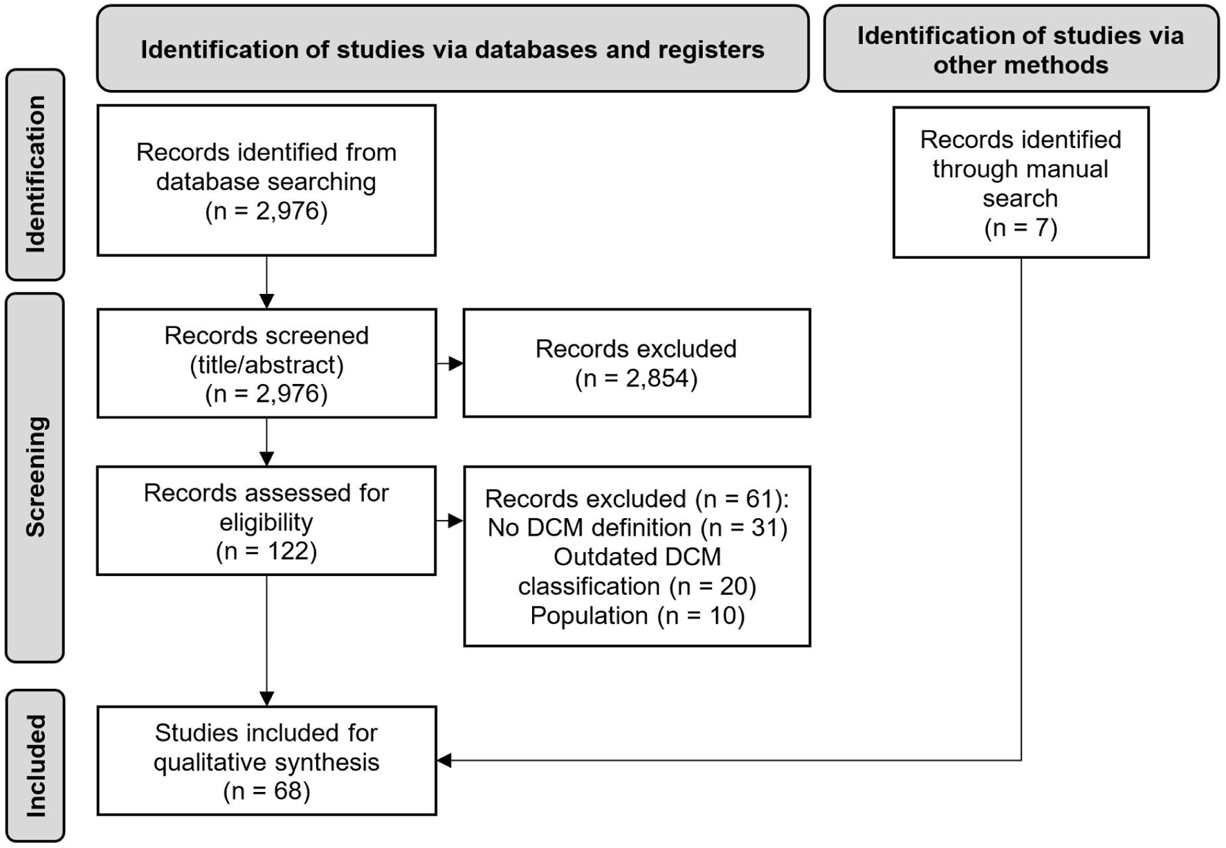
Figure 1. PRISMA diagram. n: number of records.
| Cardiology Research, ISSN 1923-2829 print, 1923-2837 online, Open Access |
| Article copyright, the authors; Journal compilation copyright, Cardiol Res and Elmer Press Inc |
| Journal website https://www.cardiologyres.org |
Review
Volume 15, Number 5, October 2024, pages 319-329
Diverse Concepts in Definitions of Dilated Cardiomyopathy: Theory and Practice
Figure
Tables
| ESC | AHA | |||
|---|---|---|---|---|
| AHA: American Heart Association; DCM: dilated cardiomyopathy; ESC: European Society of Cardiology. | ||||
| Cardiomyopathies | Primary cardiomyopathies | |||
| Hypertrophic cardiomyopathy, DCM, arrhythmogenic right ventricular cardiomyopathy, restrictive cardiomyopathy, unclassified | Genetic | Mixed | Acquired | |
| Familial/genetic | Non-familial/non-genetic | Hypertrophic cardiomyopathy | DCM | Inflammatory (myocarditis) |
| Disease subtype | Disease subtype | Arrhythmogenic right ventricular cardiomyopathy/dysplasia | Restrictive | Stress-provoked (“tako-tsubo”) |
| Unidentified gene defect | Idiopathic | Glycogen storage | - | Peripartum |
| Conduction defects | - | Tachycardia induced | ||
| Mitochondrial myopathies | - | Infants of insulin-dependent mothers | ||
| Ion channel disorders | - | |||
| Reference | Definition of dilated cardiomyopathy |
|---|---|
| DCM: dilated cardiomyopathy; HT: hypertension; LV: left ventricular; LVEF: left ventricular ejection fraction; NICM: non-ischemic cardiomyopathy. | |
| Calderon-Dominguez et al (2021) [34] | “… the presence of left ventricular dilatation and systolic dysfunction in the absence of abnormal loading conditions or coronary artery disease sufficient to cause global systolic impairment.” |
| Costa et al (2021) [36] | “DCM was defined as either LVEF levels below 50% and/or left ventricular end-diastolic diameter larger than 56 mm.” |
| Diez-Lopez et al (2022) [37] | “… left ventricular (LV) chamber enlargement and systolic dysfunction in the absence of coronary artery disease.” |
| Haas et al (2022) [42] | “Idiopathic DCM is defined as the presence of both left ventricular (LV) enlargement and systolic dysfunction but without evidence of ischemic or known causes. Specifically, diagnostic criteria for idiopathic DCM include the presence of (1) LVEF < 50% and (2) left ventricular enlargement defined by echo-derived left ventricular end-diastolic dimension (≥ 95th percentile for gender/height).” |
| Hamshere et al (2015) [45] | “… diagnosis of non-ischemic DCM with no secondary cause found, an LVEF of, 45% (assessed by echocardiography at referral), symptoms classed as New York Heart Association (NYHA) 2 or greater and on optimal medical treatment (established for at least 6 months).” |
| Kimura et al (2021) [46] | “DCM was defined by the presence of left ventricular dilation [left ventricular end-diastolic dimension (LVEDD) > 55 mm, or indexed LVEDD > 33 mm/m2 (men) or 32 mm/m2 (women)] and LVEF < 50%, in the absence of severe systemic arterial HT, coronary artery disease, primary valvular heart disease, or secondary cardiac muscle disease caused by any known systemic condition, as determined by endomyocardial biopsy.” |
| Merlo et al (2022) [49] | “NICM was defined as presence of LVEF < 50% in the absence of significant coronary artery disease, primary valve disease, congenital heart disease, tachy-induced cardiomyopathy or acute myocarditis.” |