Figures
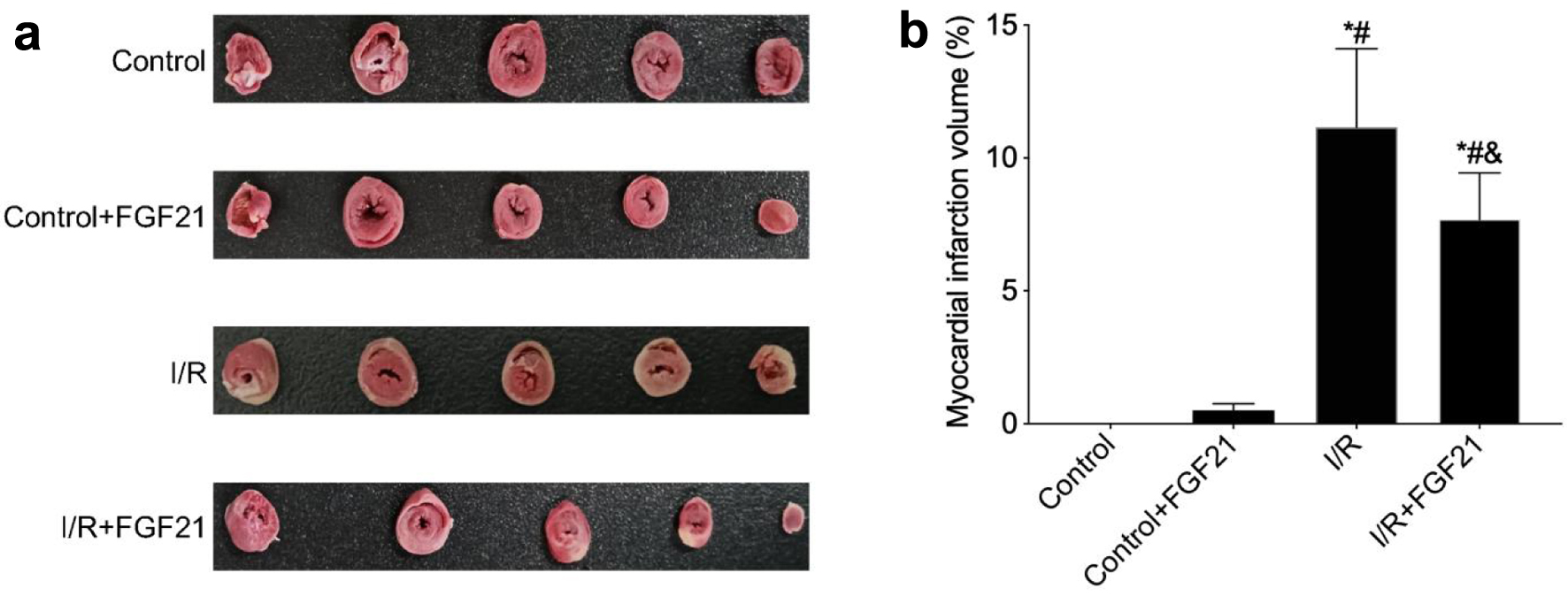
Figure 1. FGF21 inhibited I/R injury in myocardial tissue (TTC staining). (a) TTC staining of myocardial tissue. (b) Myocardial infarction volume calculated based on TTC staining. *P < 0.05 versus control group; #P < 0.05 versus control + FGF21 group; &P < 0.05 versus I/R group. FGF21: fibroblast growth factor 21; I/R: ischemia/reperfusion; TTC: 2,3,5-triphenyl tetrazolium chloride.
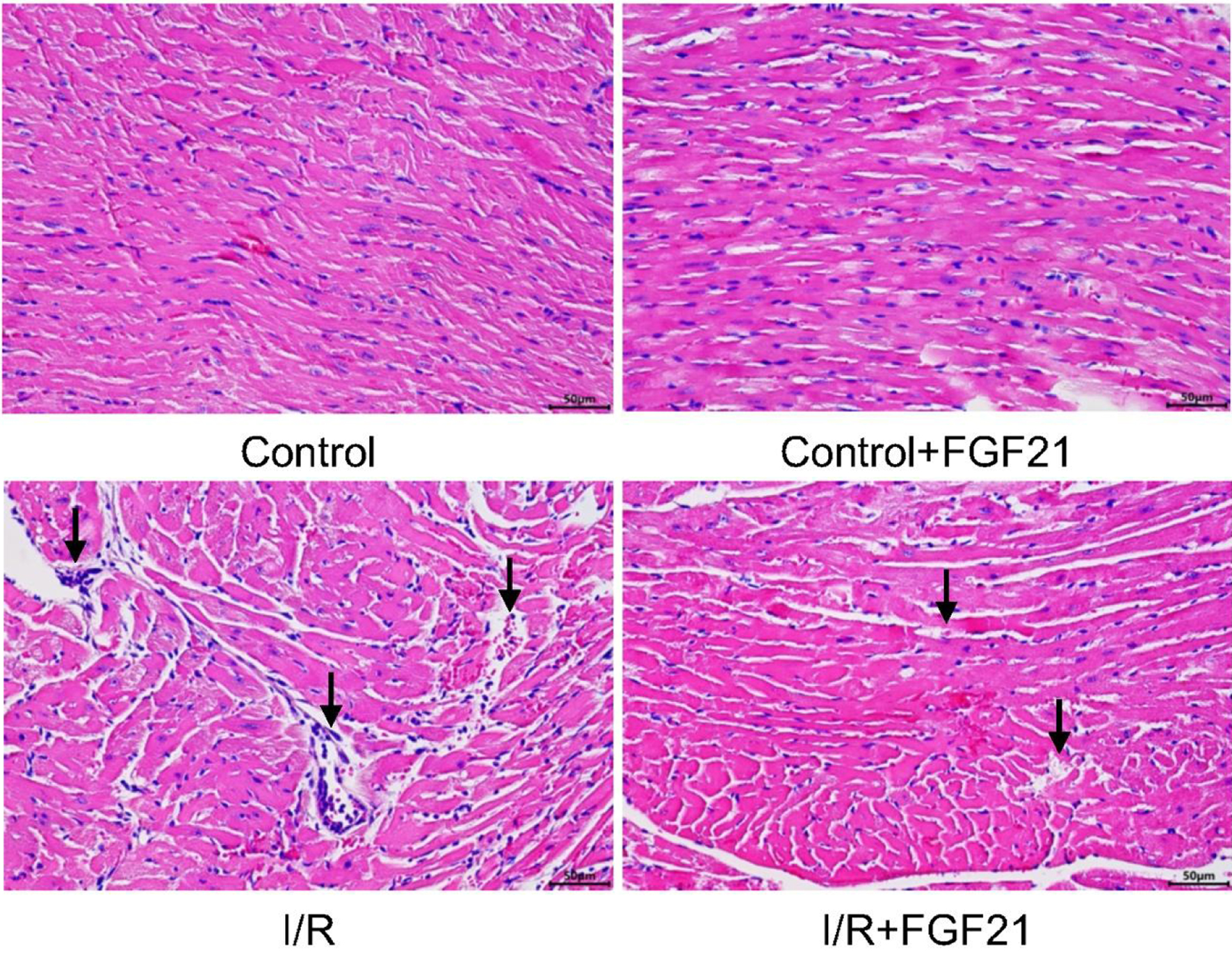
Figure 2. Myocardial tissue after myocardial I/R injury in mice pretreated with FGF21 (H&E staining, × 400). The black arrows represent obvious pathological changes in myocardial tissue. FGF21: fibroblast growth factor 21; H&E: hematoxylin and eosin; I/R: ischemia/reperfusion.
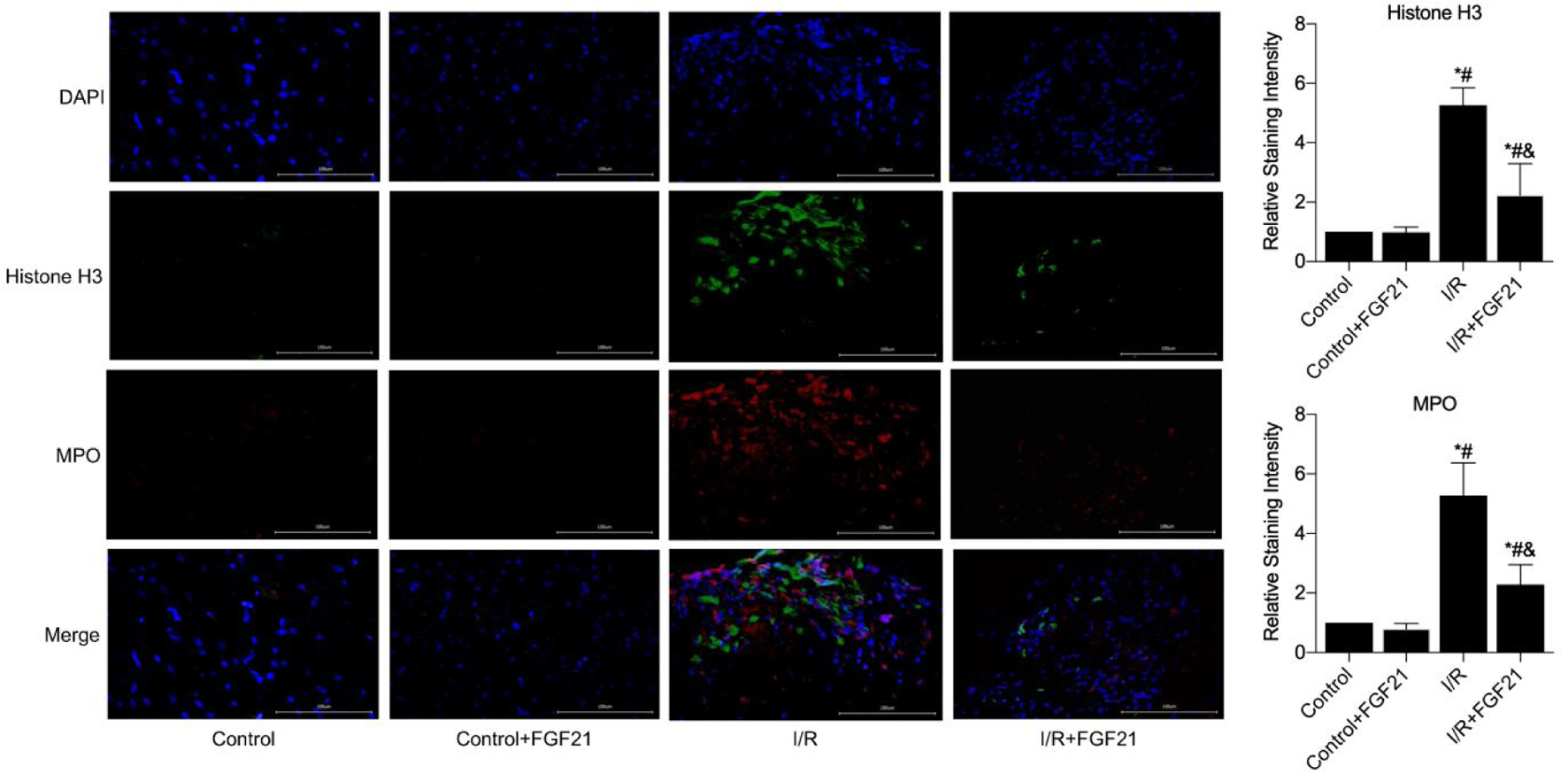
Figure 3. NETs formation in myocardial necrotic tissues was detected after the establishment of a myocardial I/R injury model in FGF21 pretreated mice (immunofluorescence, × 400). *P < 0.05 versus control group; #P < 0.05 versus control + FGF21 group; &P < 0.05 versus I/R group. FGF21: fibroblast growth factor 21; I/R: ischemia/reperfusion; NETs: neutrophil extracellular traps.
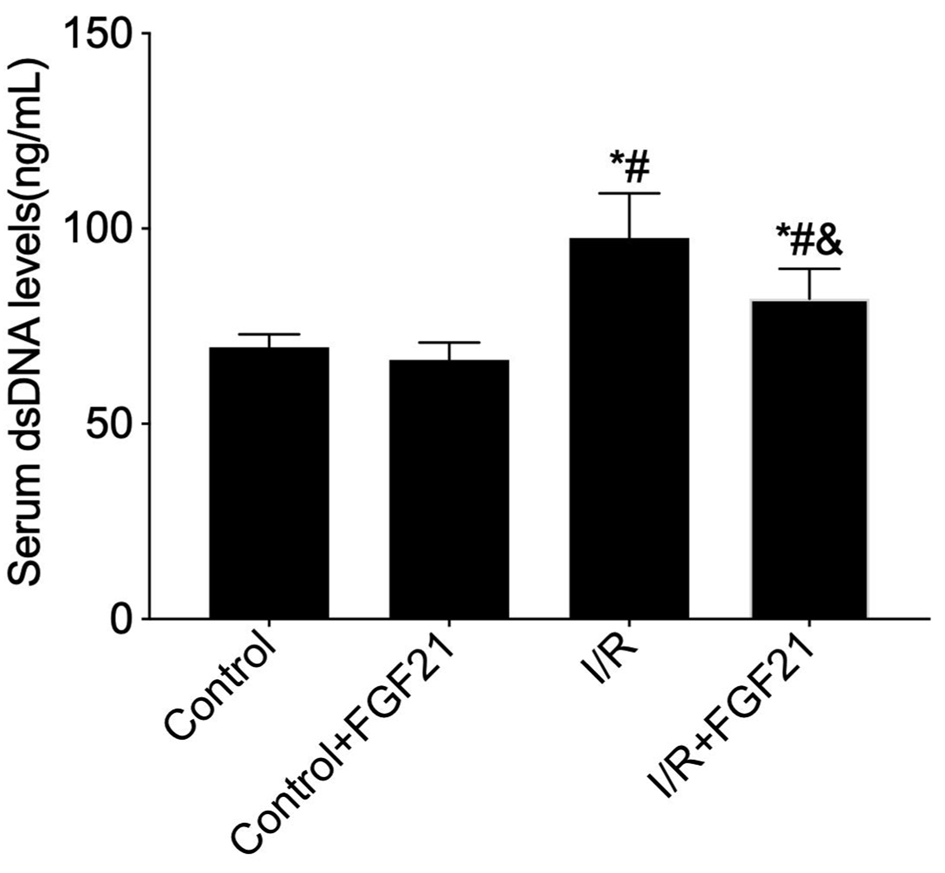
Figure 4. Measurement of peripheral serum dsDNA levels in FGF21 pretreated mice using the PicoGreen assay. *P < 0.05 versus control group; #P < 0.05 versus control + FGF21 group; &P < 0.05 versus I/R group. FGF21: fibroblast growth factor 21; I/R: ischemia/reperfusion.
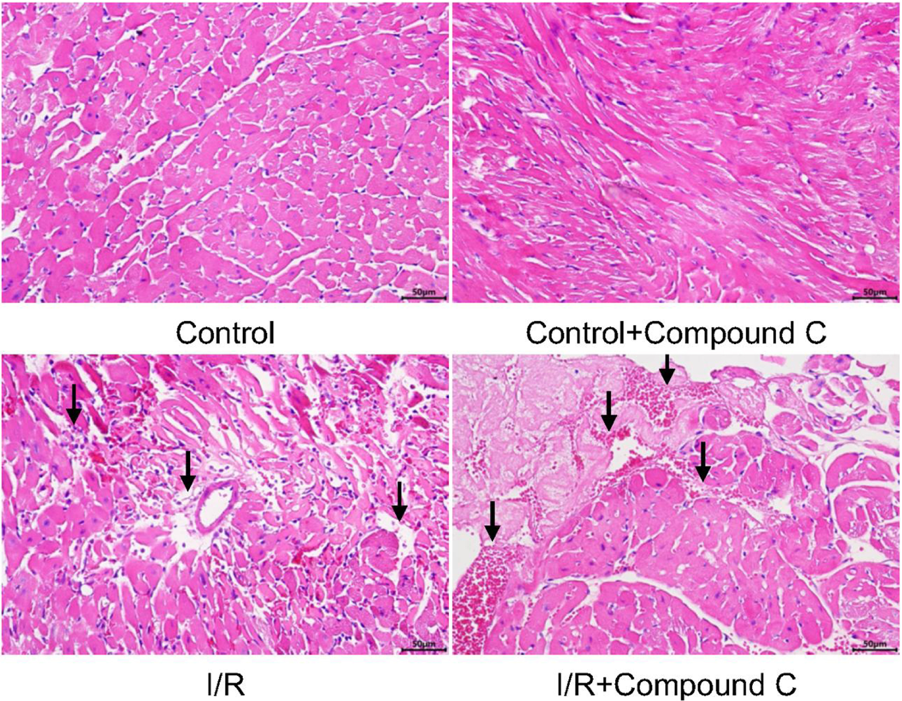
Figure 5. Myocardial tissue after myocardial I/R injury in mice pretreated with AMPK inhibitor (H&E staining, × 400). The black arrows represent obvious pathological changes in myocardial tissue. AMPK: AMP-activated protein kinase; H&E: hematoxylin and eosin; I/R: ischemia/reperfusion.
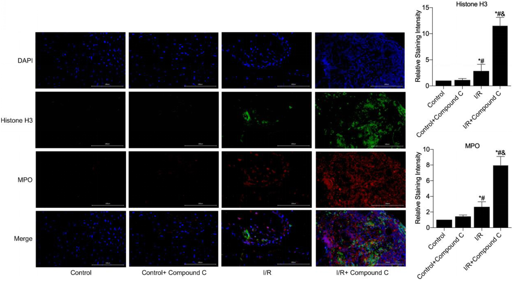
Figure 6. NETs formation in myocardial necrotic tissues was detected after the establishment of a myocardial I/R injury model in compound C pretreated mice (immunofluorescence, × 400). *P < 0.05 versus control group; #P < 0.05 versus control + FGF21 group; &P < 0.05 versus I/R group. FGF21: fibroblast growth factor 21; I/R: ischemia/reperfusion; NETs: neutrophil extracellular traps.

Figure 7. Measurement of peripheral serum dsDNA levels in compound C pretreated mice using the PicoGreen assay. *P < 0.05 versus control group; #P < 0.05 versus control + FGF21 group; &P < 0.05 versus I/R group. FGF21: fibroblast growth factor 21; I/R: ischemia/reperfusion.
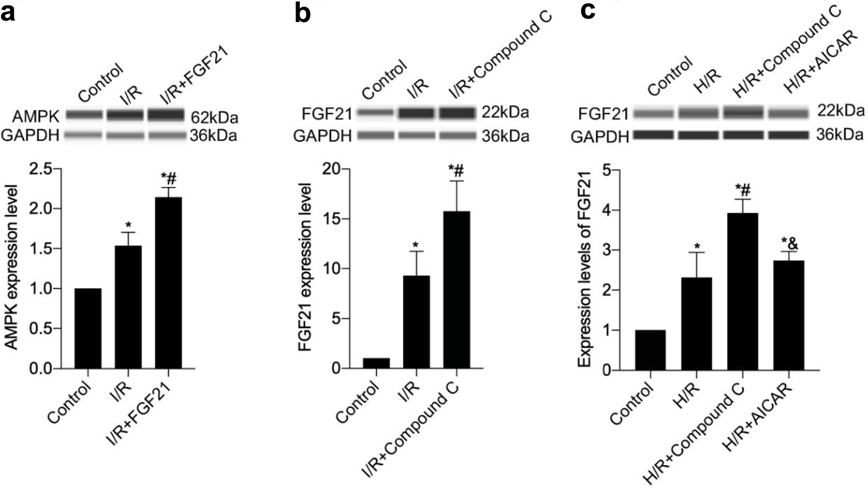
Figure 8. The interaction of FGF21 and AMPK was detected in a mouse model of myocardial I/R injury or an H/R model of H9c2 cells. (a) AMPK protein expression levels were measured in mice preinjected with FGF21. (b) FGF21 protein expression levels were measured in mice preinjected with AMPK inhibitor compound C. (c) FGF21 protein expression levels were measured in H9c2 cells pretreated with compound C, an inhibitor of AMPK, or with AICAR, an agonist. *P < 0.05 versus control; #P < 0.05 versus I/R group; &P < 0.05 versus H/R + compound C group. AMPK: AMP-activated protein kinase; FGF21: fibroblast growth factor 21; H/R: hypoxia/reoxygenation; I/R: ischemia/reperfusion.
Figure 9. AMPK inhibited H/R injury in H9c2 cells in a time-dependent manner. *P < 0.05 versus control; #P < 0.05 versus H/R group; &P < 0.05 versus H/R + compound C group. AMPK: AMP-activated protein kinase; H/R: hypoxia/reoxygenation.