Cardiology Research, ISSN 1923-2829 print, 1923-2837 online, Open Access
Article copyright, the authors; Journal compilation copyright, Cardiol Res and Elmer Press Inc
Journal website https://www.cardiologyres.org

Case Report

Volume 5, Number 6, December 2014, pages 188-190


Purulent Pericarditis Leading to Constriction

Figures

Figure 1.
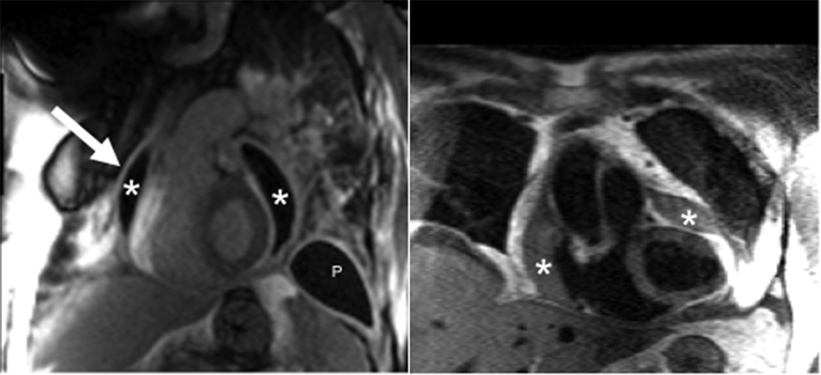
Figure 1. Post-gadolinium inversion recovery sequence with steady state free precession readout demonstrates a grossly thickened pericardium (arrow) accompanied by a loculated pericardial effusion (*). Pleural thickening with effusion (P) is also present. Enhancement of the pleura and pericardium is consistent with significant fibrosis. Double inversion T1-weighted turbo spin echo sequence also demonstrates the findings of a loculated pericardial effusion.
Figure 2.
Figure 2. Simultaneous right and left ventricular recordings showing discordant ventricular filling during inspiration, consistent with constrictive physiology.
Figure 3.
Figure 3. Intraoperative photo reveals a severely thickened parietal pericardium.