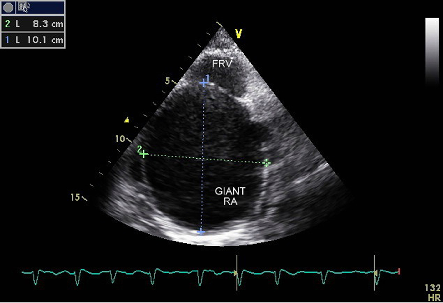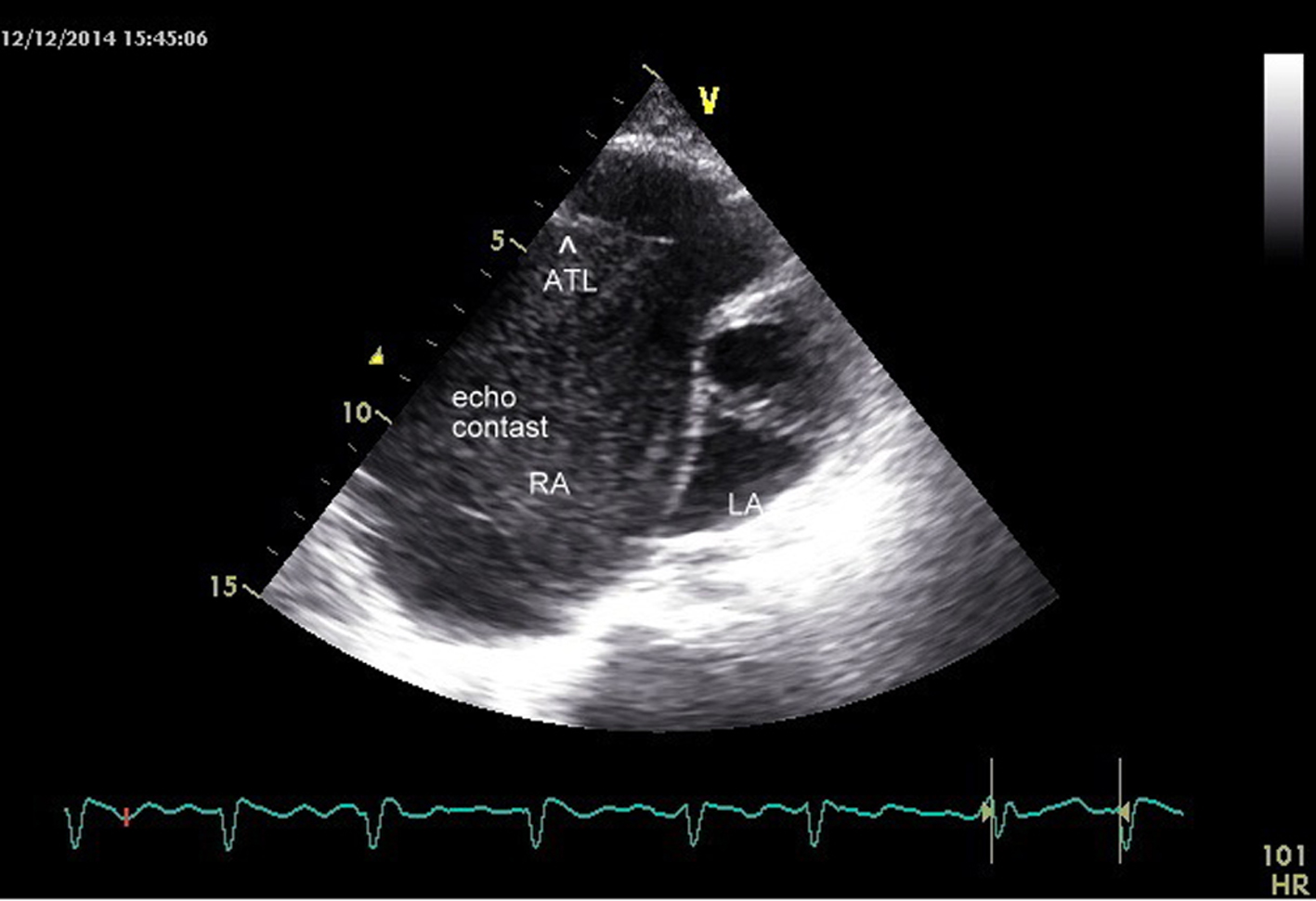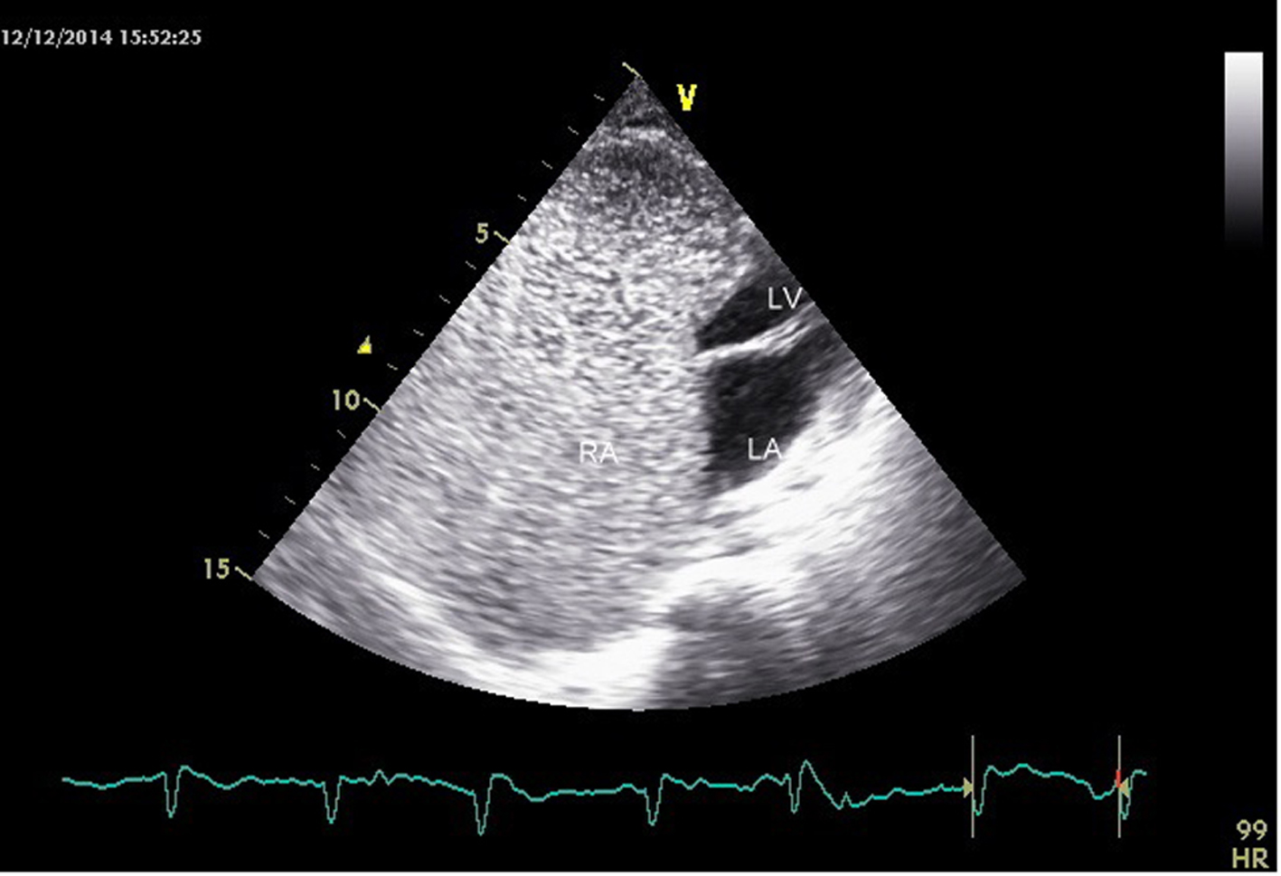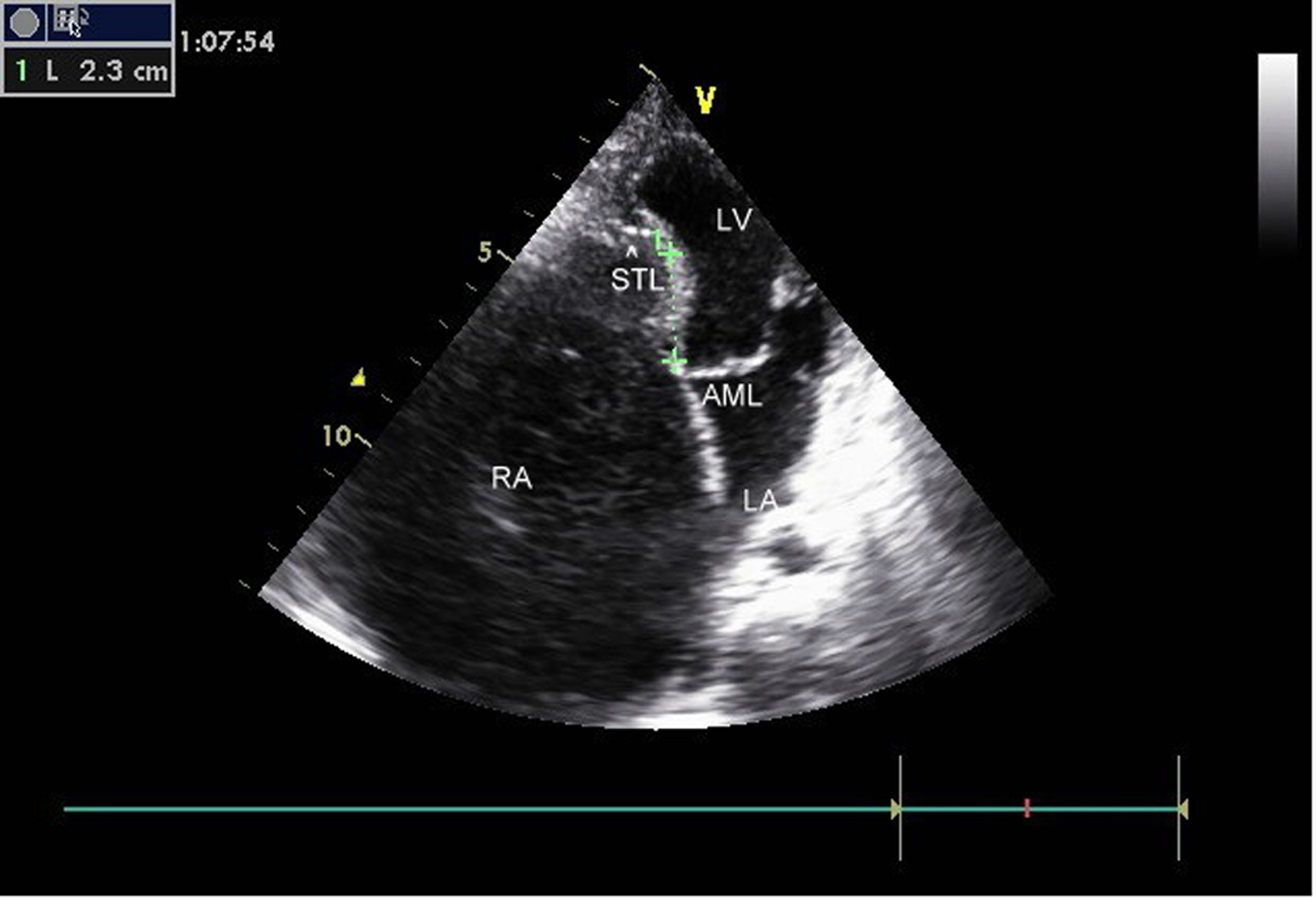
Figure 1. Ebstein anomaly, 2.3 cm apical displacement of septal leaflet (arrow head showing STL, septal leaflet of TV) and massively dilated RA.
| Cardiology Research, ISSN 1923-2829 print, 1923-2837 online, Open Access |
| Article copyright, the authors; Journal compilation copyright, Cardiol Res and Elmer Press Inc |
| Journal website https://www.cardiologyres.org |
Case Report
Volume 6, Number 4-5, October 2015, pages 319-323
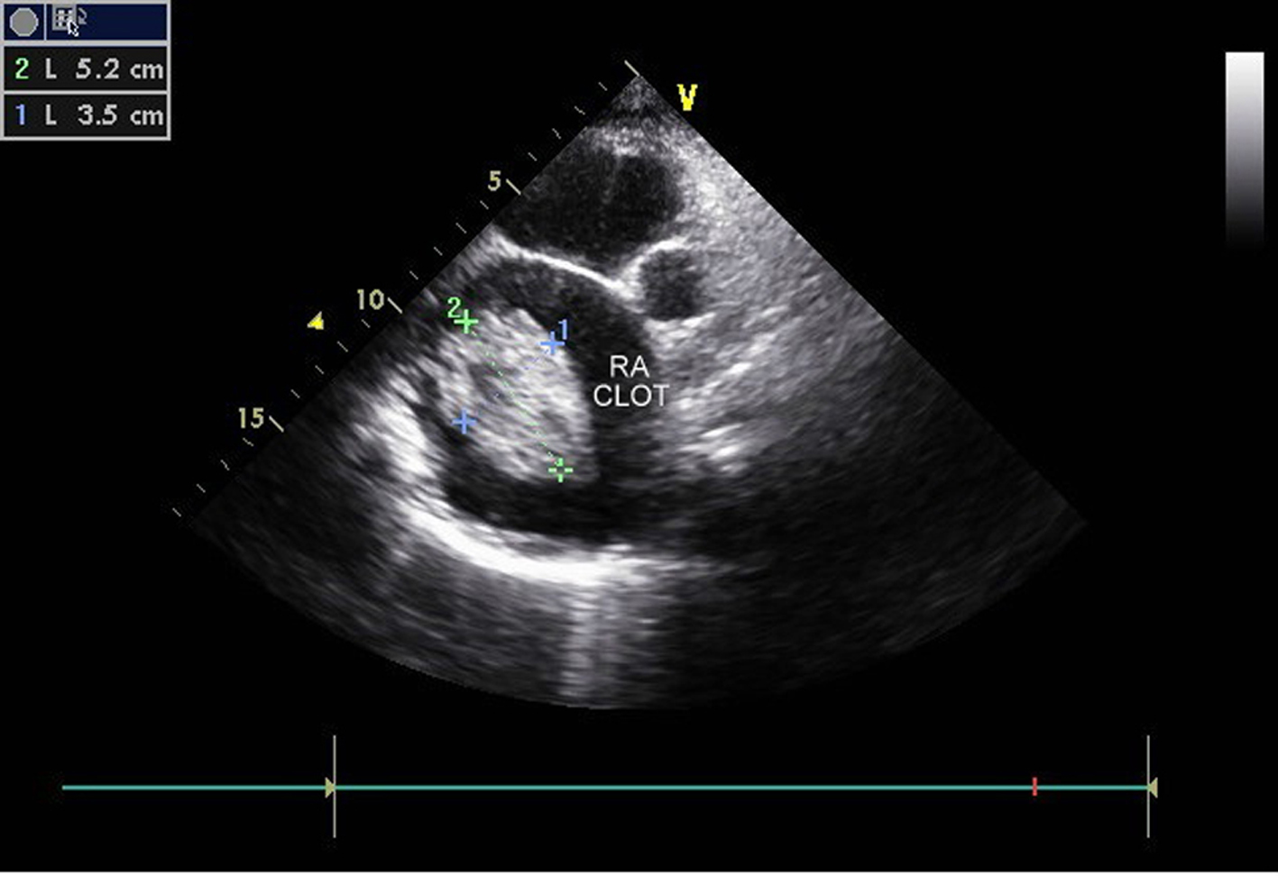
Ebstein Anomaly With Right Atrial Clot
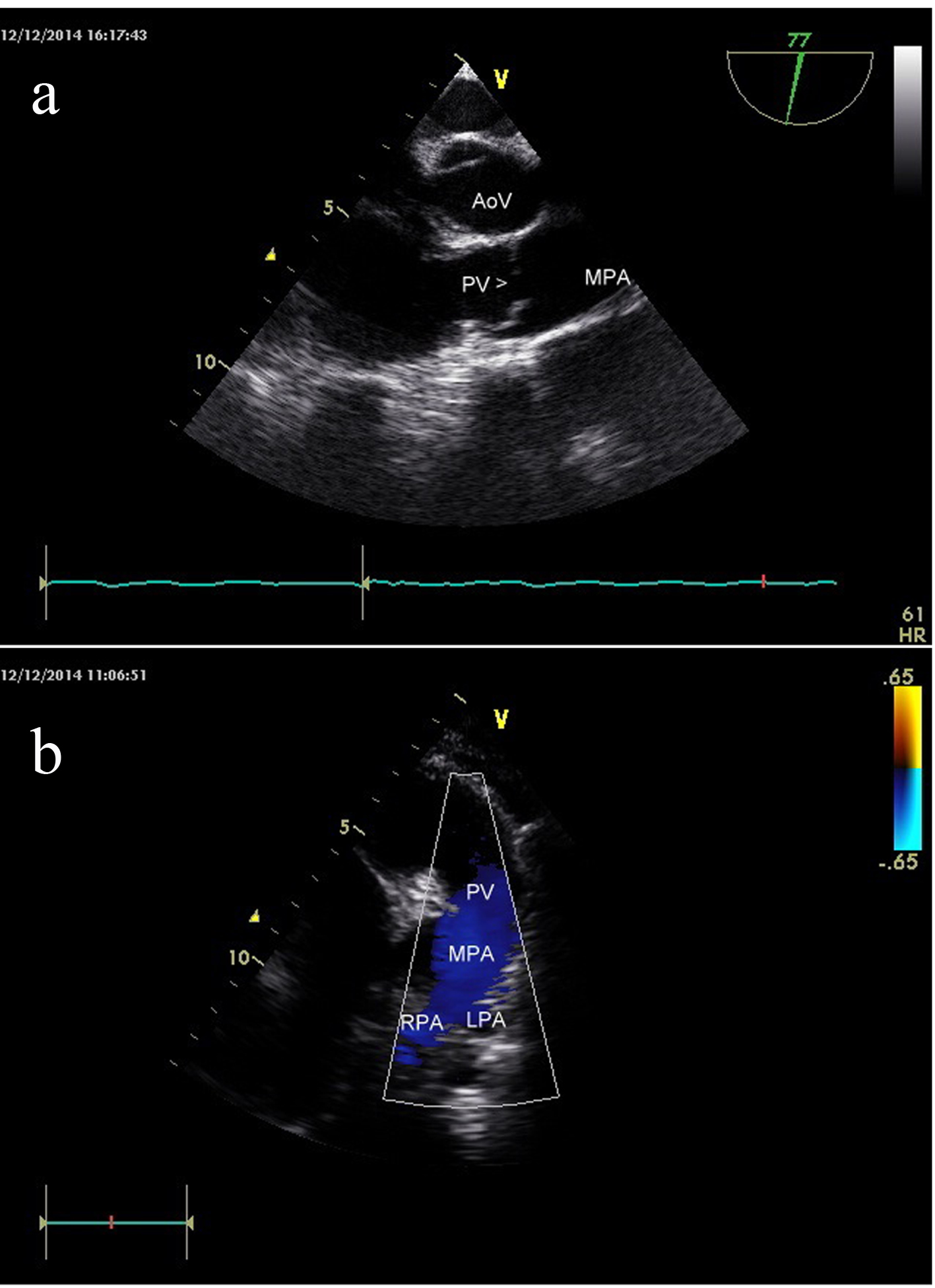
Figures