Cardiology Research, ISSN 1923-2829 print, 1923-2837 online, Open Access
Article copyright, the authors; Journal compilation copyright, Cardiol Res and Elmer Press Inc
Journal website https://www.cardiologyres.org

Case Report

Volume 7, Number 3, June 2016, pages 119-121


A Sudden Change of Heart: A Case of Rapidly Reversed Stress Cardiomyopathy in a Critically Ill Patient

Figures

Figure 1.
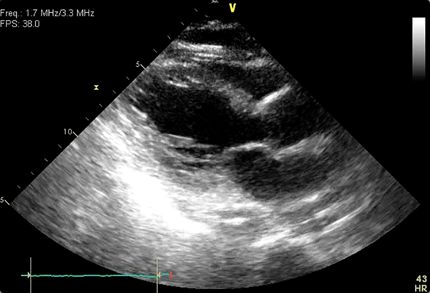
Figure 1. Parasternal long axis view during systole. Ballooning of the apical segments of the left ventricle is apparent.
Figure 2.
Figure 2. Post-arrest coronary angiogram. There is right dominant circulation. The coronary arteries are diffusely constricted. The left main is angiographically normal. The left anterior descending artery has minimal disease (A). The left circumflex has an ostial 30% stenosis (B). The right coronary artery has minimal disease, and supplies the posterior descending artery (C).
Figure 3.
Figure 3. ECG taken 12 h after cardiac arrest. Note the low voltages, inferior and lateral precordial lead ST segment depressions, and the prolonged QT interval.