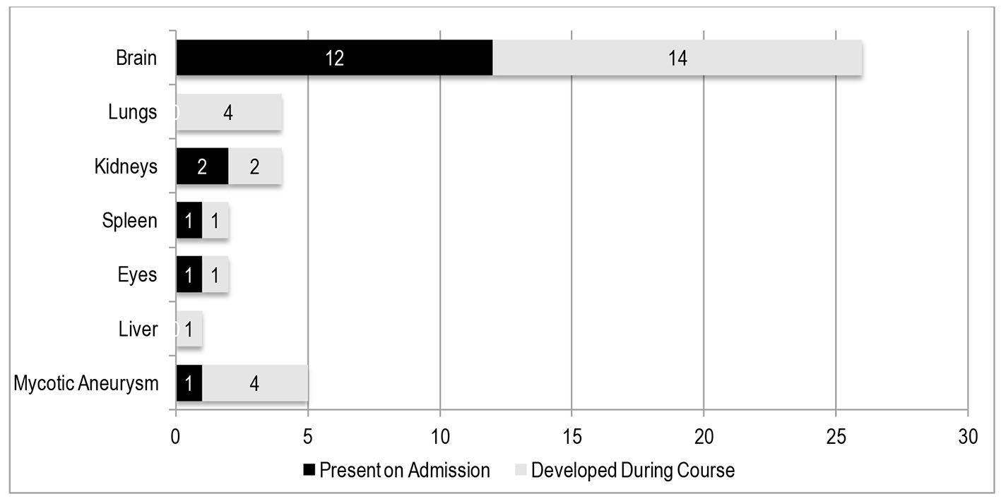
Cardiology Research, ISSN 1923-2829 print, 1923-2837 online, Open Access
Article copyright, the authors; Journal compilation copyright, Cardiol Res and Elmer Press Inc
Journal website https://www.cardiologyres.org

Original Article

Volume 7, Number 4, August 2016, pages 130-139


Prediction of Symptomatic Embolism in Filipinos With Infective Endocarditis Using the Embolic Risk French Calculator

Figures

Figure 1.
Figure 1. Distribution of cardioembolic events. Embolic events were present on admission (prior to initiation of antibiotic therapy) in 17 patients. The primary outcome (i.e. embolic events during/after initiation of antibiotic therapy) occurred in 25 patients.
Figure 2.
Figure 2. Causes of death. Mortality occurred in 25 patients (33%) prior to the 28-day observation period.

Tables

Table 1. Definition of Variables
 
Variables collectedDefinition/description
Presence of minor emboli on admissionAny minor embolic events present on admission, before the initiation of antibiotic therapy, such as Janeway lesions, Roth spots, splinter hemorrhages, petechial hemorrhages, and subconjunctival hemorrhage.
Presence of major emboli on admissionAny major embolic events present on admission, before the initiation of antibiotic therapy, further defined below.
Organism on blood cultureOrganism present on at least two sets of blood cultures
Presence of atrial fibrillationPersistent or transient atrial fibrillation documented on 12-lead electrocardiogram or on a cardiac monitor.
Length of vegetationVegetation length measured in various planes and the maximal length was recorded. For multiple vegetations, the largest length was used for analysis. A vegetation length ≥ 10 mm is considered to be a large vegetation.
Area of vegetationComputed by the longest length multiplied by the widest width of a single vegetation. For multiple vegetations, the individual computed areas were added. An area of ≥ 18 mm2 is considered to be a large vegetation.
Ejection fractionEchocardiographically derived by the Teicholz method. For studies with wall motion abnormalities, the EF derived by Simpson’s method was recorded.
Significant valvular regurgitationDefined as a “severe” regurgitant lesion (e.g., severe MR, severe TR).
Location of vegetationLeft-sided vegetations pertain to those located on the left side of the heart, including the MV, AV, endocardium of the LA or LV, or aorta. Right-sided vegetations pertain to those located on the right side, including the TV, PV, endocardium of the RA or RV, or the pulmonary artery.
Prosthetic valve vegetationIncludes mechanical or bioprosthetic valves.


Table 2. Definition of Major Embolic Events
 
Major embolic eventsDefinition of terms
Arterial emboliSudden interruption of blood flow to an organ system due to an embolus, presumed by the physician to be from infective endocarditis. This complication is manifested by symptoms of end organ damage. For example, CNS emboli - manifestations would include stroke-like symptoms/neurologic deficits, documented by cranial imaging (CT or MRI). Other examples would include splenic, renal, peripheral, hepatic, retinal, or mesenteric emboli.
Intracranial hemorrhageBleeding into the CNS presumed to be due to a CNS emboli, which is not attributable to any other etiology such as hypertension or trauma.
Pulmonary infarctsLung injury due to interruption of blood flow to the lungs due to a (pulmonary) embolus. Manifestations may include sudden onset dyspnea, hemoptysis or pulmonary hemorrhage, or hypoxemia not attributable to any other cause. This may be documented by chest radiograph or CT/MRI.
Mycotic aneurysmsAneurysm arising from bacterial infection of the arterial wall. Manifestations would include end-organ damage attributable to the involved system (i.e. neurologic deficits for CNS mycotic aneurysm, sudden abdominal pain for aortic mycotic aneurysms).


Table 3. Baseline Clinical Profile Stratified by Outcome (Development of Embolus)
 
VariableTotal (n = 87) (%)With embolus+ (n = 25) (%)Without embolus (n = 62) (%)P-value
+Development major embolism after initiation of antibiotic therapy. **Minor embolic events include splinter hemorrhage, Janeway lesions or conjunctival hemorrhage.
Age (mean ± SD)39.90 (13.98)36.68 (11.40)41.19 (14.78)0.175
  > 55 years old15 (17.24)2 (8.00)13 (20.97)0.147
  ≤ 55 years old72 (82.76)23 (92.00)49 (79.00)
Male sex49 (56.32)16 (64.00)33 (53.23)0.359
Presence of minor emboli on admission**44 (50.57)15 (60.00)29 (46.77)0.264
Mean duration of illness in weeks7.21 (7.75)5.00 (5.41)8.10 (8.38)0.092
  < 6 weeks (acute-subacute)57 (65.51)19 (76.00)38 (61.29)0.191
  ≥ 6 weeks (chronic)30 (34.48)6 (24.00)24 (38.71)
Organism on blood culture
  Staphylococcus spp.13 (14.94)7 (28.00)6 (9.68)0.030
  Streptococcus viridans21 (24.14)7 (28.00)14 (22.58)0.593
  Gram negative bacteria11 (12.64)3 (12.00)8 (12.90)0.909
  Culture negative40 (45.98)8 (32.00)32 (51.61)0.097


Table 4. Distribution According to Predisposing Cardiac Condition by Echocardiography
 
Predisposing cardiac conditionFrequency (n = 87) (%)
Valvular heart disease (rheumatic and non-rheumatic)59 (68%)
  Mitral stenosis25
  Mitral valve prolapse14
  Aortic stenosis9
Congenital heart disease (CHD)15 (17%)
  Ventricular septal defect8
  Patent ductus arteriosus3
  Repaired CHD2
  Pulmonic stenosis1
Prosthetic valve endocarditis5 (7%)
Unknown or no underlying predisposition8 (11%)


Table 5. Components of the French Risk Calculator
 
VariableTotal (n = 87) (%)With embolus+ (n = 25) (%)Without embolus (n = 62) (%)P-value
+Major embolic events included any arterial emboli, intracranial hemorrhage, pulmonary infarcts, or mycotic aneurysms. *P value < 0.05.
Mean age (SD)39.86 (13.95)36.72 (11.39)41.13 (14.75)0.183
Presence of diabetes mellitus18 (20.69)6 (24.00)12 (19.35)0.628
Major emboli prior to admission18 (20.69)11 (44.00)7 (11.29)0.001*
Atrial fibrillation23 (26.44)12 (48.00)11 (17.74)0.004*
Length of vegetation on 2D echocardiogram
  < 10 mm51 (58.62)7 (28.00)44 (70.97)< 0.001*
  ≥ 10 mm35 (40.23)18 (72.00)17 (27.42)< 0.001*
Staphylococcus aureus on blood culture8 (9.20)6 (24.00)2 (3.23)0.002*
Mean score on French calculator (SD)5.13 (4.33)8.68 (5.75)3.69 (2.48)< 0.001*


Table 6. Echocardiographic Profile Stratified by Outcome (Development of Embolus)
 
VariableTotal (n = 87) (%)With embolus+ (n = 25) (%)Without embolus (n = 62) (%)P-value
+Development major embolism after initiation of antibiotic therapy. ++Left atrial volume index. +++Vegetation area was computed by multiplying the longest length with the widest width. *P-value < 0.05.
Overall cardiac structure and function
  Mean ejection fraction (SD)60.92 (11.57)60.32 (12.70)61.16 (11.18)0.761
    Ejection Fraction < 45%11 (12.64)5 (20.00)6 (9.68)0.190
  Mean LAVI (mL/m2)++44.56 (29.84)41.40 (25.76)45.84 (31.44)0.533
    LAVI > 34 mL/m254 (62.07)17 (68.00)37 (59.68)0.469
  Significant valvular regurgitation39 (44.83)12 (48.00)27 (43.55)0.706
Number of vegetations
  0 vegetation5 (5.75)2 (8.00)3 (4.84)0.566
  1 vegetation64 (73.56)16 (64.00)48 (77.42)0.199
  > 1 vegetation18 (20.69)7 (28.00)11 (17.74)0.285
Size and characteristics of the vegetation
  Vegetation length (mean ± SD)8.45 (6.11)10.60 (6.49)7.58 (5.78)0.036*
    Vegetation length ≥ 10 mm35 (40.23)16 (64.00)19 (30.65)0.004*
    Vegetation length > 15 mm14 (16.09)6 (24.00)8 (12.90)0.202
    Vegetation area (mean ± SD)+++70.40 (85.22)112.72 (100.97)53.33 (72.09)0.003*
  Vegetation area ≥ 18 mm250 (57.47)21 (84.00)29 (46.77)0.001*
  Mobile vegetation61 (70.11)21 (84.00)40 (64.52)0.072
  Presence of abscess6 (6.90)2 (8.00)4 (6.45)0.796
Location of vegetation
  Left-sided vegetation79 (90.80)21 (84.00)58 (93.55)0.163
  Right-sided vegetation10 (11.49)5 (20.00)5 (8.06)0.114
Valvular involvement
  Mitral location56 (64.37)17 (68.00)39 (62.90)0.653
  Aortic location21 (24.14)7 (28.00)14 (22.58)0.593
  Tricuspid/pulmonic location8 (9.20)5 (20.00)3 (4.84)0.027*
  Multiple valves19 (21.84)8 (32.00)11 (17.74)0.145
  Prosthetic valves3 (3.45)1 (4.00)2 (3.23)0.858
  No valves (endocardium)10 (11.49)3 (12.00)7 (11.29)0.925


Table 7. Univariate Analysis to Predict Development of Embolic Events during Admission
 
VariableRelative risk95% CIP-value
+Included in the final model of multivariate analysis. *High risk embolic events using the embolic risk French calculator were set at > 7% risk at 1 month (see references).
High risk for embolic events (> 7%)*20.275.95 - 69.05< 0.001*
Age > 550.330.07 - 1.570.163
Diabetes mellitus1.040.32 - 3.340.945
Minor embolism prior to therapy1.710.66 - 4.380.266
Major embolism prior to therapy8.622.73 - 27.23< 0.001*
Atrial fibrillation3.851.41 - 10.520.009*
Ejection fraction < 45%2.330.64 - 8.490.199
Left-sided vegetation0.360.08 - 1.580.176
More than one vegetation1.800.61 - 5.360.289
Vegetation length ≥ 10 mm4.021.51 - 10.710.005*
Vegetation length ≥ 15 mm2.130.65 - 6.940.209
Area of vegetation ≥ 18 mm25.971.84 - 19.440.003*
Staphylococcus aureus on culture3.631.08 - 12.210.037*


Table 8. Multivariate Analysis to Predict Embolic Events
 
VariableRelative risk95% CIP-value
+Independent predictors of embolic events with P value < 0.05. *High risk embolic events using the embolic risk French calculator were set at > 7% risk at 1 month.
High risk score for embolic events (> 7%)*15.124.19 - 54.54< 0.001+
Vegetation area ≥ 18 mm26.391.55 - 26.310.010+
Major embolism prior to therapy5.181.32 - 20.350.018+


Table 9. Multiple Logistic Regression to Predict Primary and Secondary Outcomes
 
VariableEmbolic event (primary endpoint)Composite of death and embolic eventsDeath
RRP-valueRRP-valueRRP-value
*High risk score pertains to a high embolic risk score at one month, calculated using the French calculator. RR: relative risk; NS: not significant.
High risk score (> 7%)*15.12< 0.00113.56< 0.0016.20.003
Vegetation area ≥ 18 mm26.390.010NSNS
Emboli prior to admission5.180.01813.750.002NS
Ejection fraction ≤ 45%NSNS9.910.004