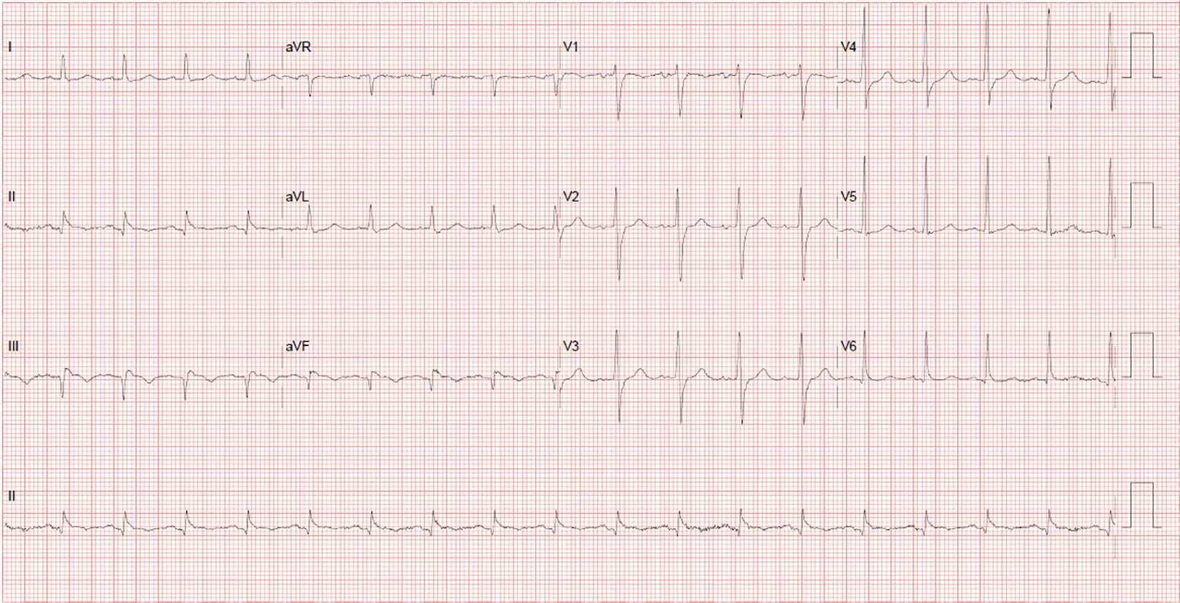
Figure 1. Q waves in the inferior leads with 2 mm ST elevations and T-wave inversions, suggestive of a recent inferior myocardial infarction.
| Cardiology Research, ISSN 1923-2829 print, 1923-2837 online, Open Access |
| Article copyright, the authors; Journal compilation copyright, Cardiol Res and Elmer Press Inc |
| Journal website https://www.cardiologyres.org |
Case Report
Volume 7, Number 5, October 2016, pages 181-184
Late Presentation of an Inferior Myocardial Infarction Complicated by Ventricular Septal Rupture: A Case Report
Figures