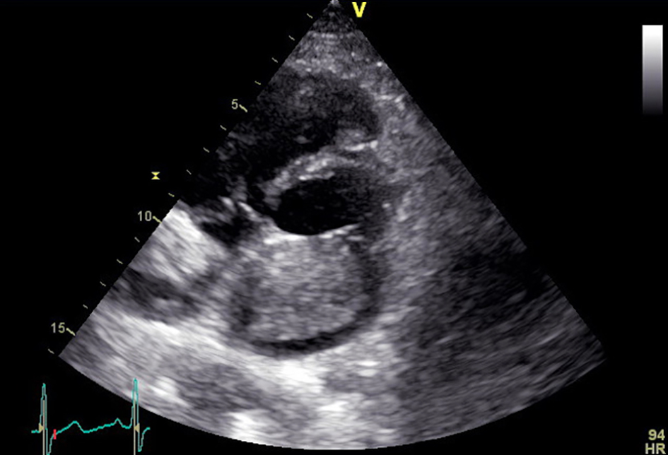
Figure 1. Long parasternal axis view of transthoracic echocardiography showing a left atrial myxoma reaching the atrial surface of the mitral valve.
| Cardiology Research, ISSN 1923-2829 print, 1923-2837 online, Open Access |
| Article copyright, the authors; Journal compilation copyright, Cardiol Res and Elmer Press Inc |
| Journal website https://www.cardiologyres.org |
Case Report
Volume 8, Number 3, June 2017, pages 128-130
Atrial Myxoma Mimicking Mitral Stenosis
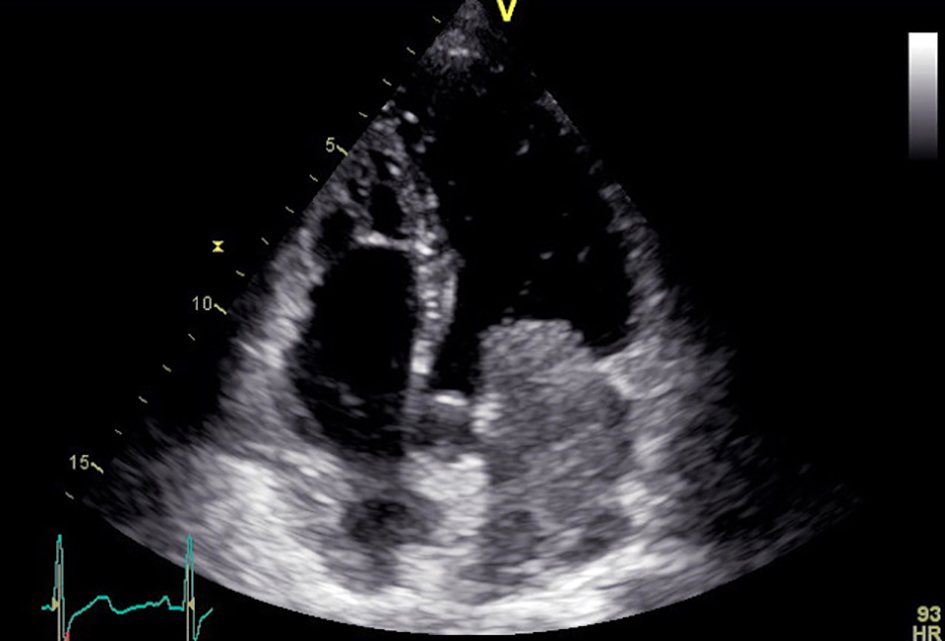
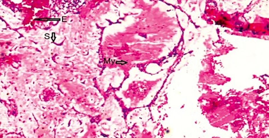
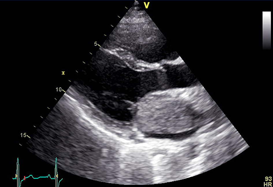
Figures