Figures
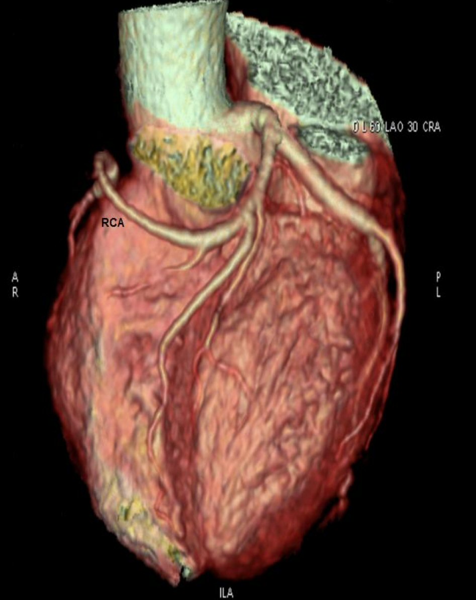
Figure 1. MSCT showing RCA coming from LAD as a continuation of septal branch.
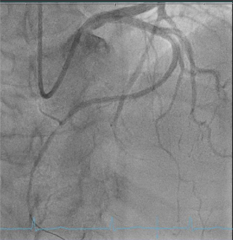
Figure 2. Coronary angiography LAO 8/CR 31 view showing RCA coming from LAD with mid segment RCA stenosis 70-90%.
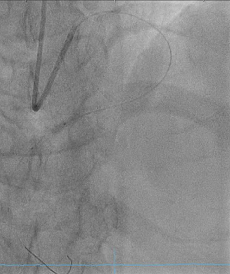
Figure 3. Coronary angiography LAO 8/CR 31 view showing mid RCA stent deployment.
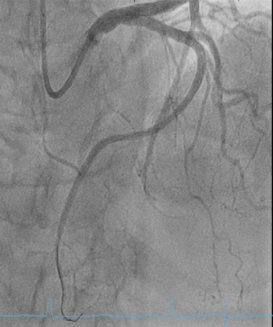
Figure 4. Coronary angiography LAO 8/CR 31 view showing good results after RCA stenting.
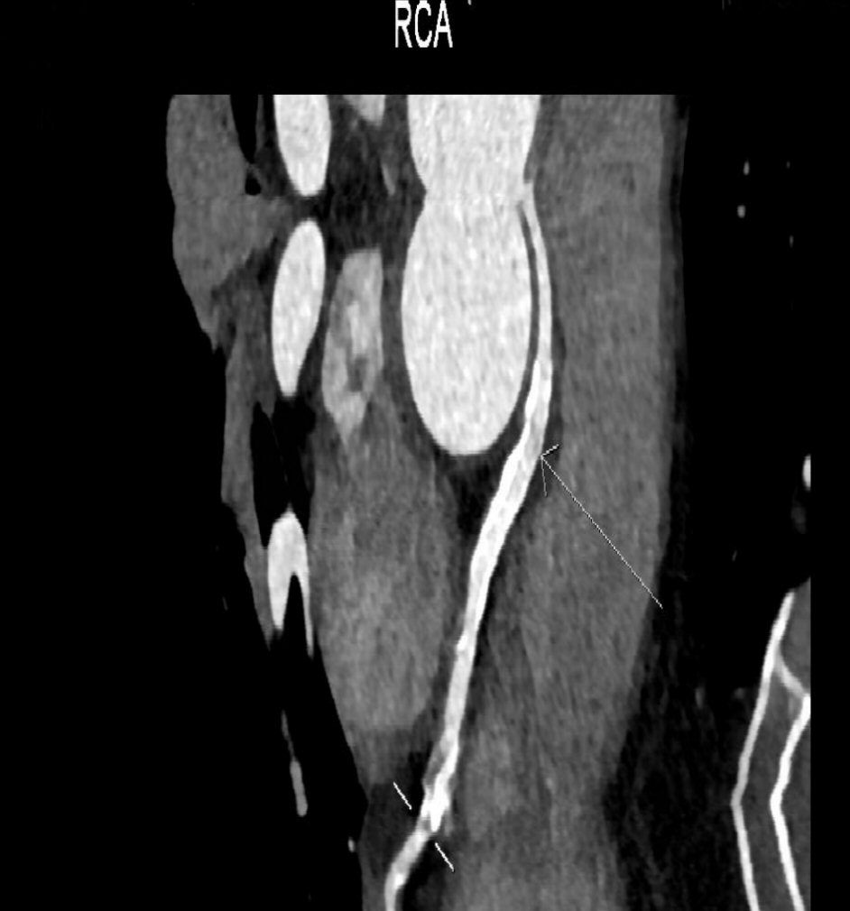
Figure 5. MSCT showing RCA coming from left main coronary artery.
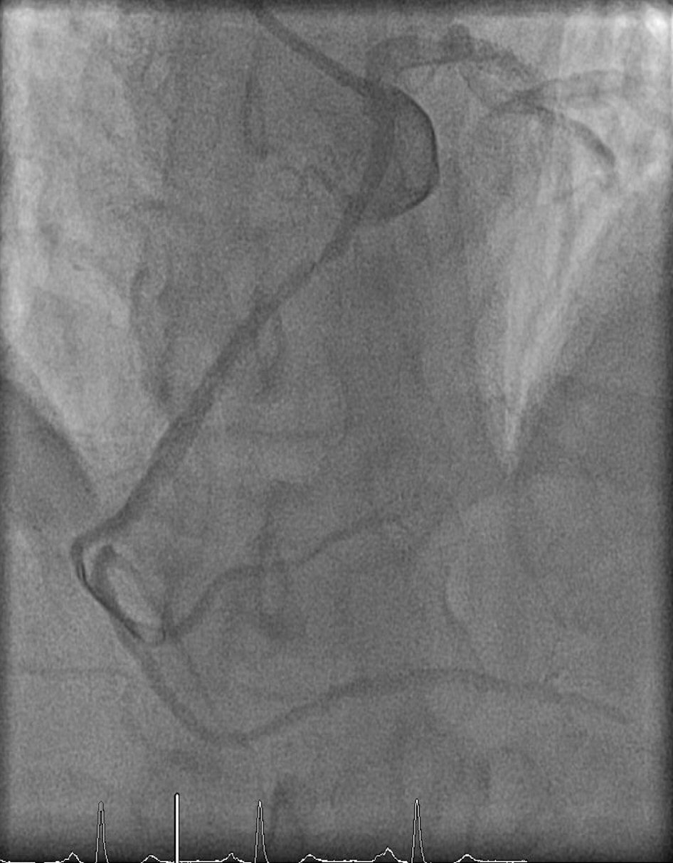
Figure 6. Coronary angiography LAO 6/CR 36 view showing RCA coming from LM with 90% in-stent restenosis at proximal segment.
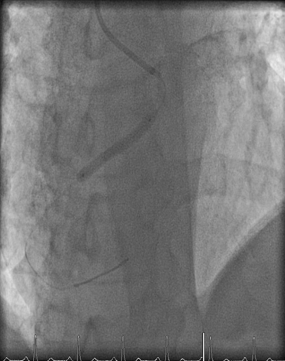
Figure 7. Coronary angiography LAO 6/CR 36 view showing proximal RCA stent deployment.
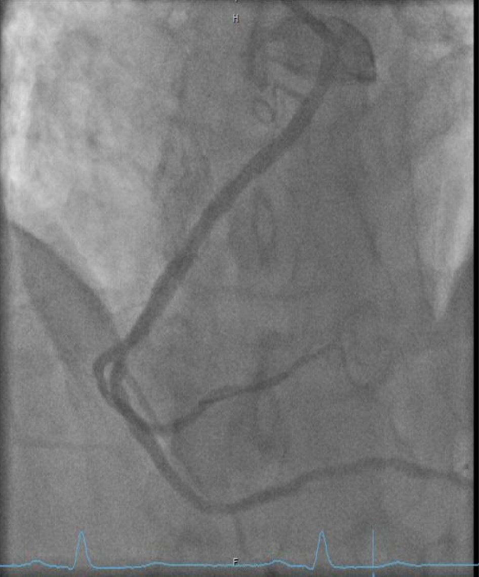
Figure 8. Coronary angiography LAO 6/CR 36 view showing good final results after proximal RCA stent deployment.
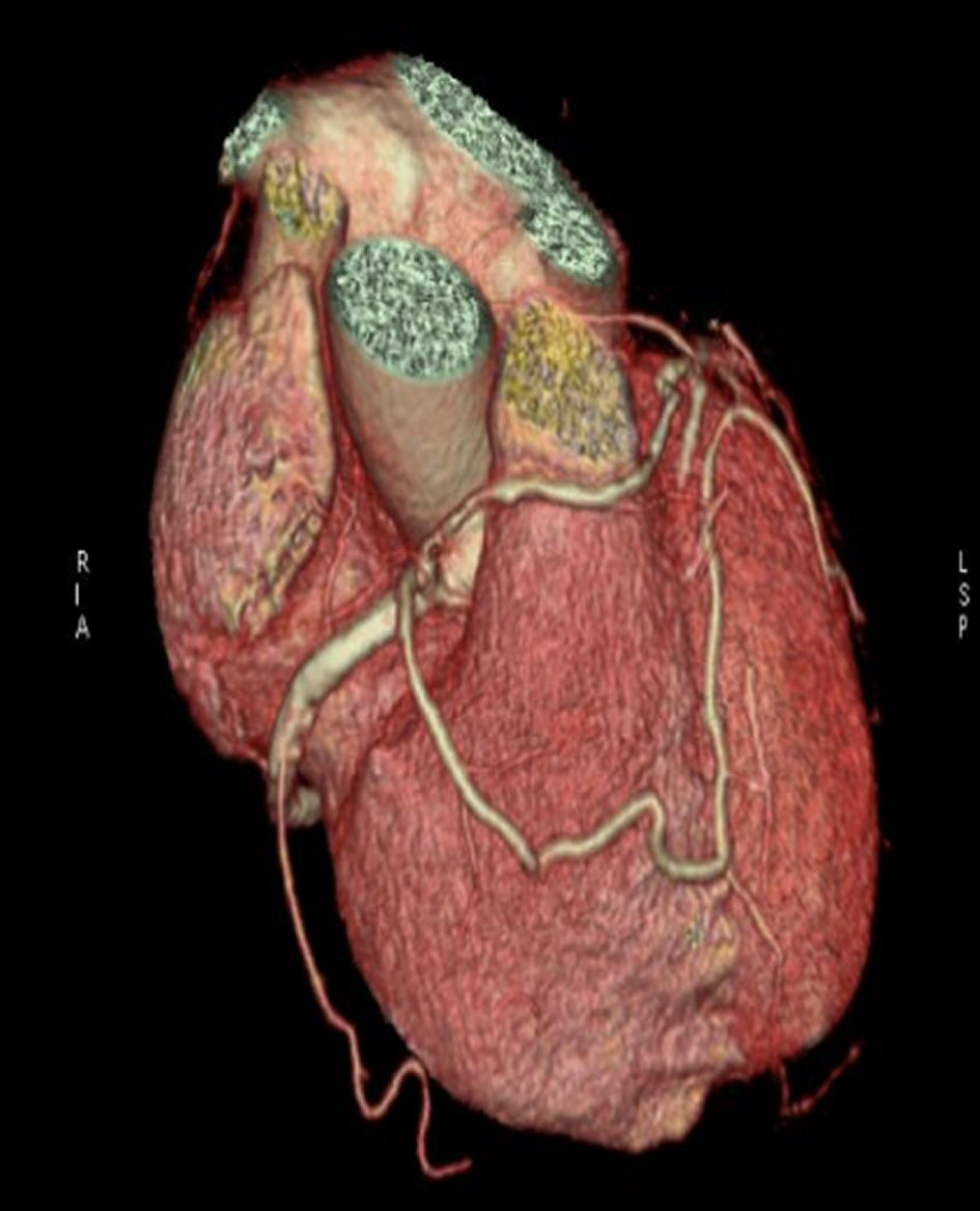
Figure 9. MSCT showing LAD and RCX coming from proximal RCA.
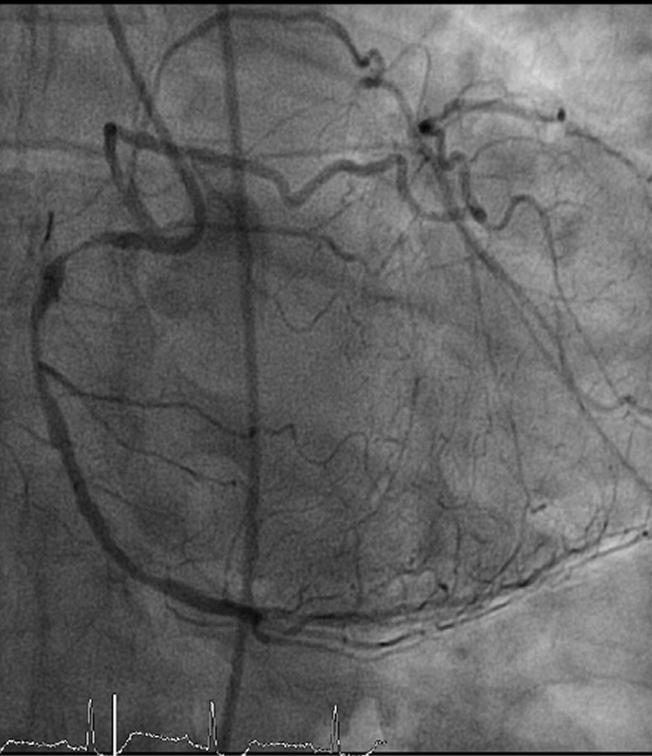
Figure 10. Coronary angiography RAO 20/CA 28 view showing LAD and LCX coming from proximal RCA with 99% stenosis at proximal and mid RCA.
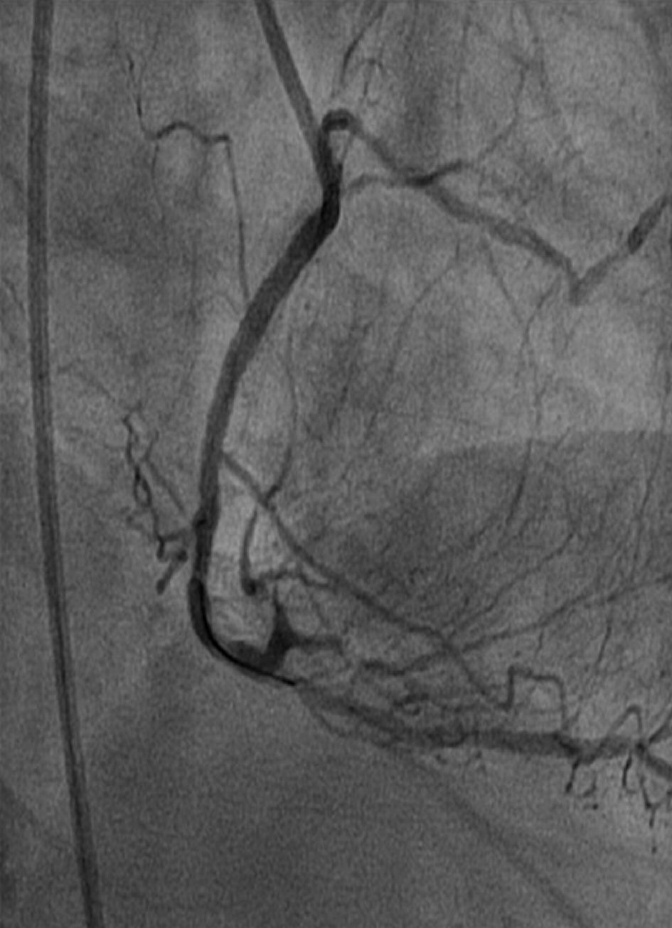
Figure 11. Coronary angiography RAO 30/CA 50 view showing good results after proximal and mid RCA stent deployment.