Figures
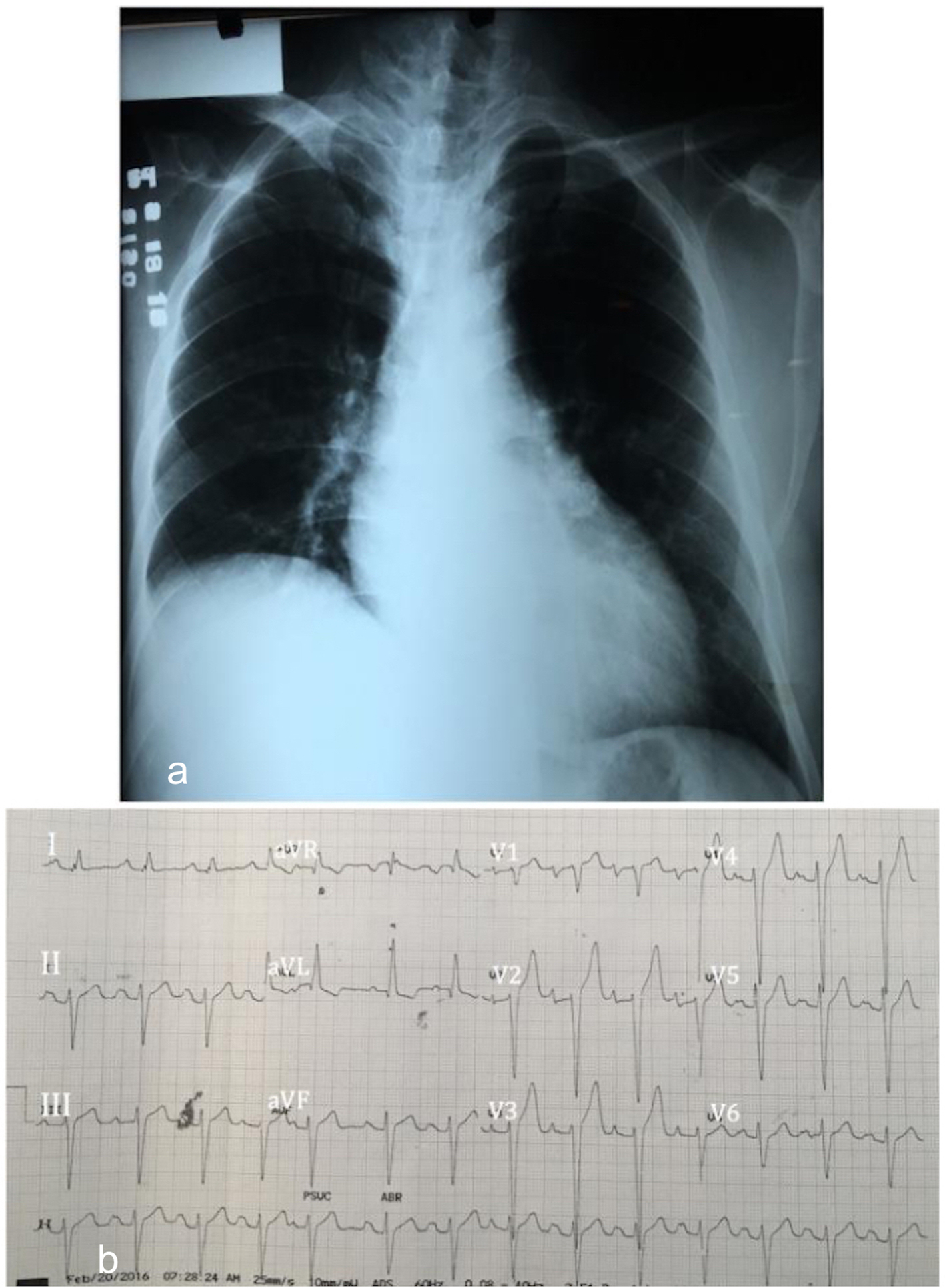
Figure 1. (a) Chest radiograph showing normal cardiac borders with loss of normal cardiac waistline suggestive of left atrial enlargement. (b) Electrocardiogram showing regular sinus rhythm, left axis deviation, left anterior fascicular block, first-degree atrioventricular block, bi-atrial abnormality and biventricular hypertrophy, isolated premature atrial complex.
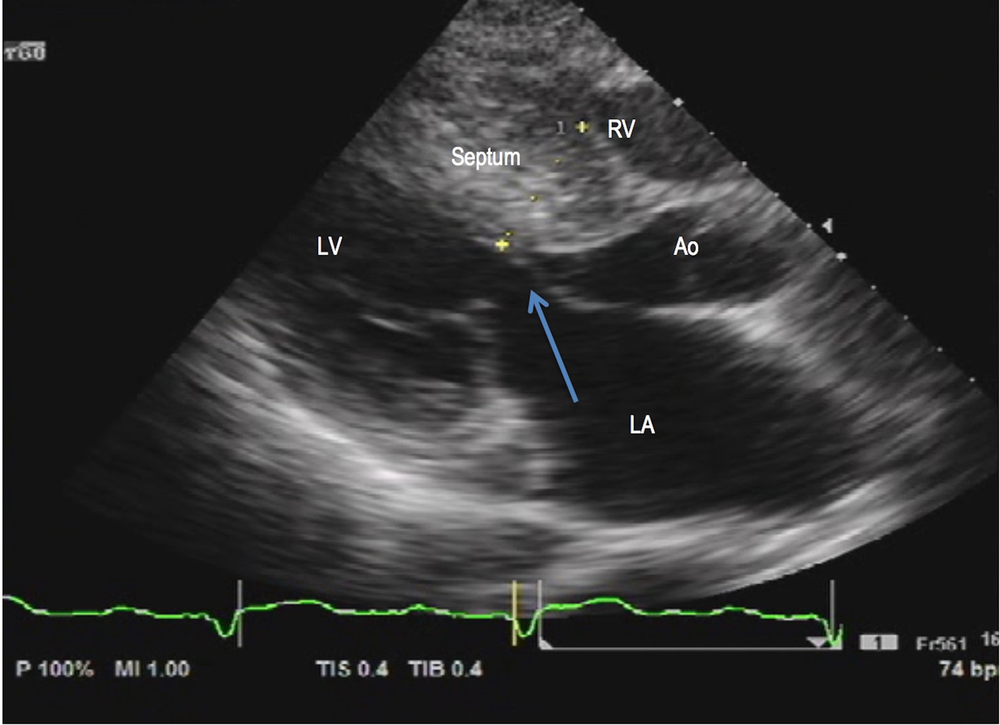
Figure 2. 2D transthoracic echocardiography. Asymmetric LV hypertrophy (septal to free wall thickness ratio of 1.6) and systolic anterior motion of the anterior mitral valve (arrow) during early systole seen on parasternal long axis view. LA: left atrium; LV: left ventricle; RV: right ventricle; Ao: aorta.
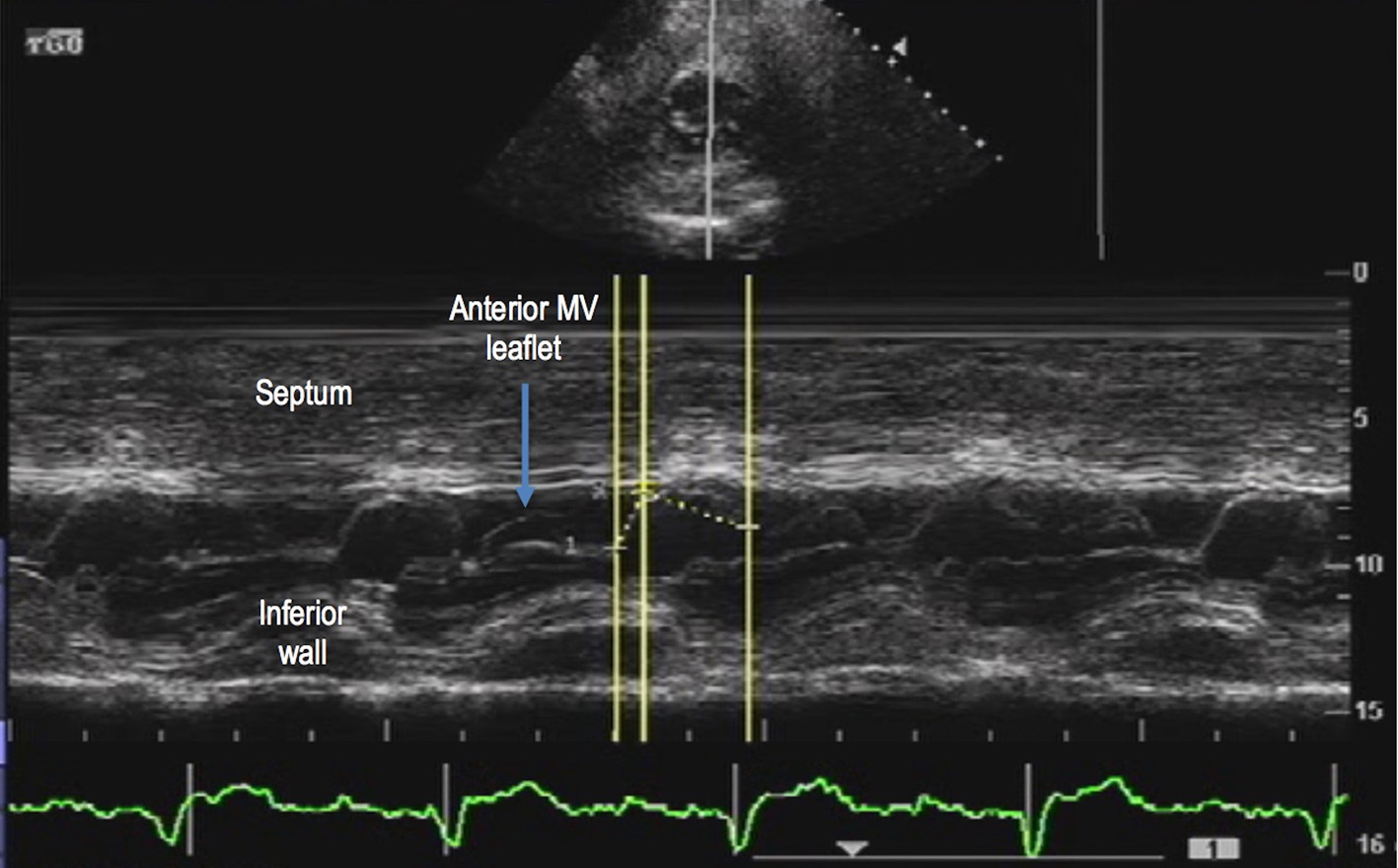
Figure 3. 2D transthoracic echocardiography. M-mode at the level of the mitral valve on parasternal short axis view confirmed systolic anterior motion (arrow) of the anterior mitral valve (MV) leaflet.
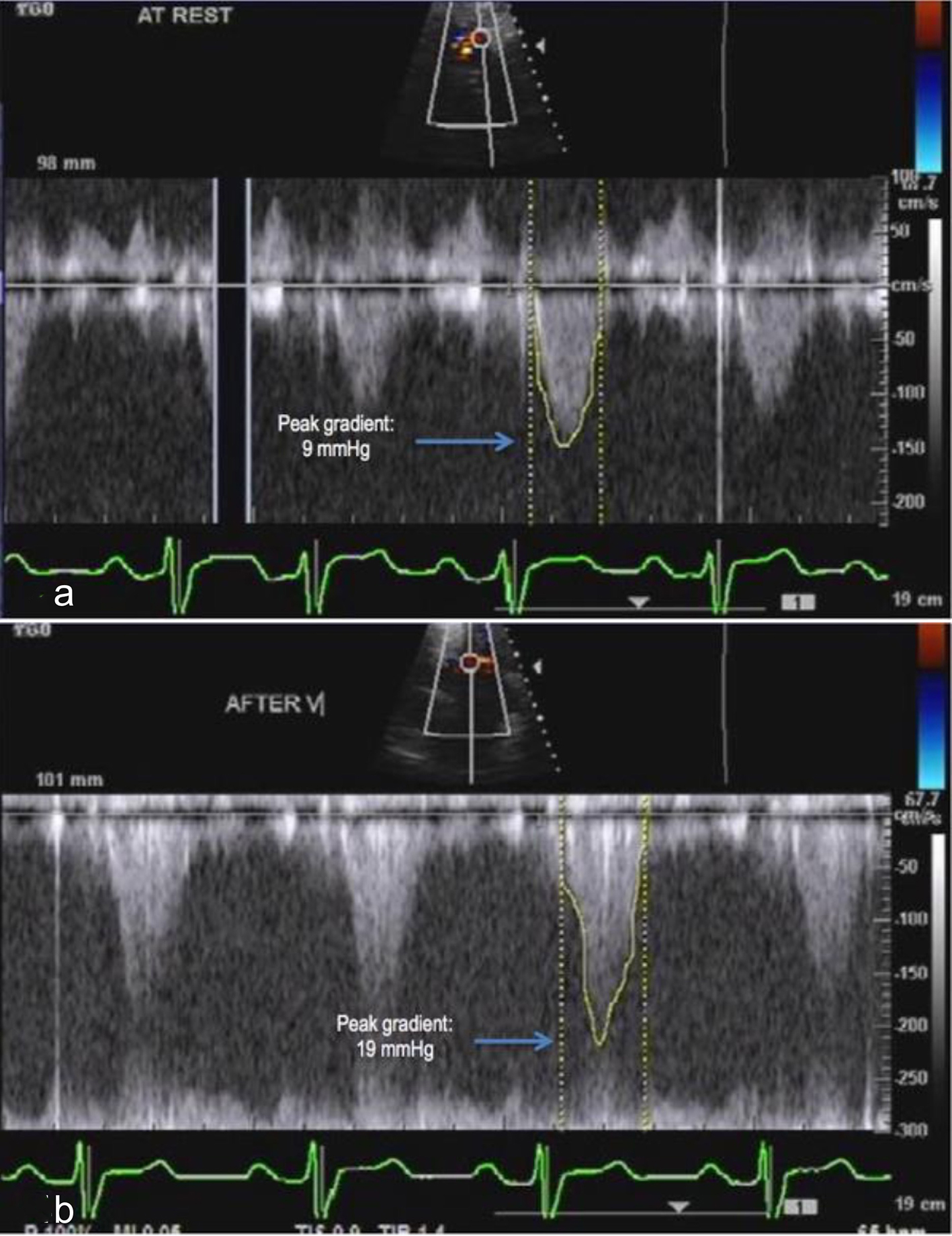
Figure 4. Continuous wave Doppler. (a) Interrogation of gradient along the LVOT revealed a peak gradient of 9 mm Hg. (b) Provocative maneuvers such as Valsalva maneuver increased the peak gradient to more than twice the gradient at rest, suggestive of some yet non-significant LVOT obstruction.
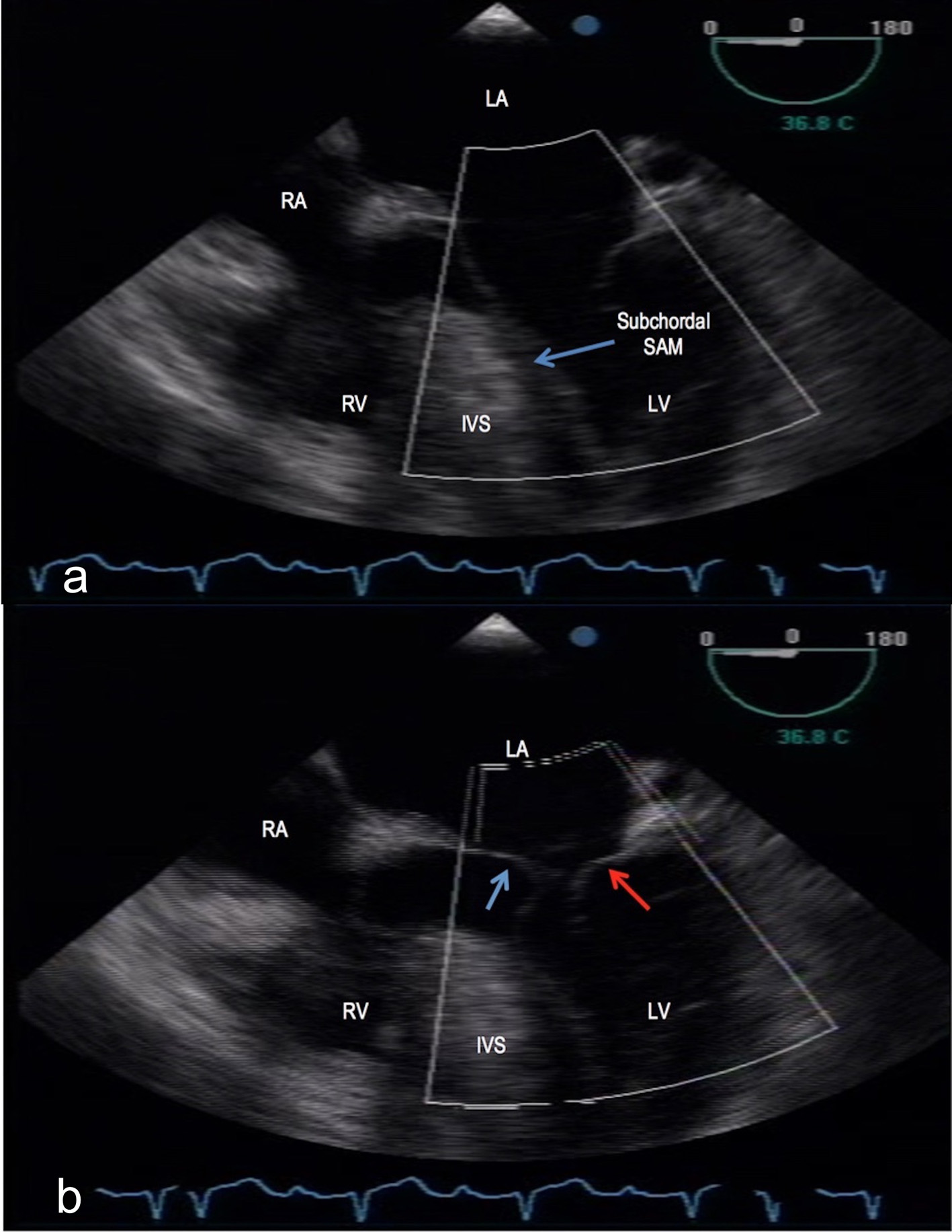
Figure 5. Transesophageal echocardiography, mid-esophageal level, 0°. (a) A redundant anterior mitral valve with the tip and subchordal apparatus causing systolic motion (arrow) towards a hypertrophied interventricular septum (IVS). (b) Thickened anterior (blue arrow) and posterior (red arrow) mitral valve leaflets with posterior displacement into the left atrium during systole indicative of mitral valve prolapse. LA: left atrium; RA: right atrium; RV: right ventricle; LV: left ventricle; SAM: systolic anterior motion; IVS: interventricular septum.
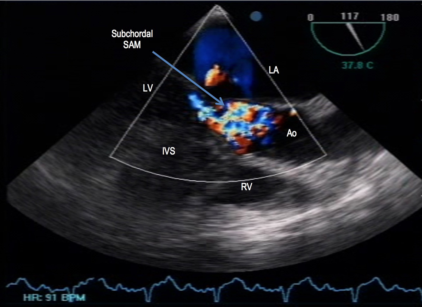
Figure 6. Transesophageal echocardiography, transgastric level, 117°. Mosaic color flow was seen at the level of the LVOT during systole, suggestive of some LVOT obstruction. LA: left atrium; RV: right ventricle; LV: left ventricle; Ao: aorta; IVS: interventricular septum; SAM: systolic anterior motion.
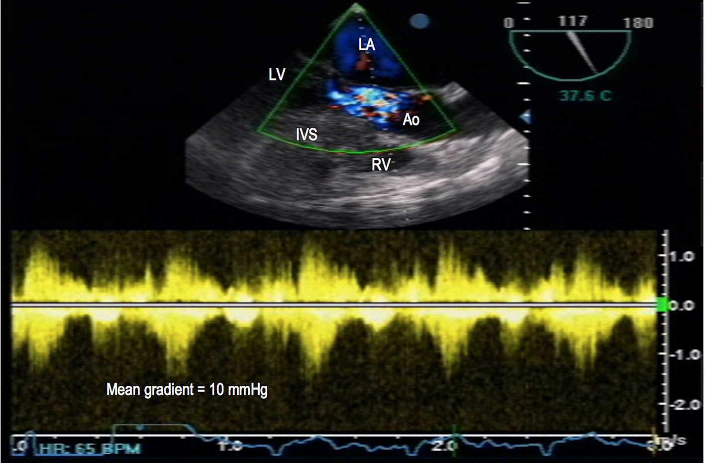
Figure 7. Transesophageal echocardiography. Continuous wave Doppler revealed a mean gradient of 10 mm Hg across the left ventricular outflow tract (LVOT). LA: left atrium; RV: right ventricle; LV: left ventricle; Ao: aorta; IVS: interventricular septum.