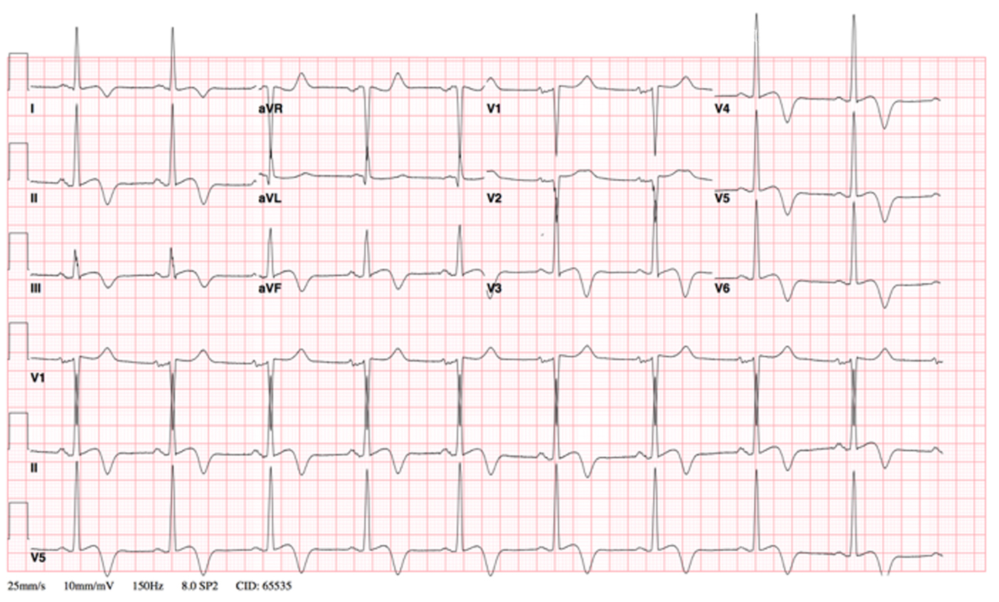
Figure 1. EKG shows normal sinus rhythm, left ventricle hypertrophy and marked T-wave inversions in leads II, III and aVF.
| Cardiology Research, ISSN 1923-2829 print, 1923-2837 online, Open Access |
| Article copyright, the authors; Journal compilation copyright, Cardiol Res and Elmer Press Inc |
| Journal website https://www.cardiologyres.org |
Case Report
Volume 8, Number 5, October 2017, pages 265-268
Apical Hypertrophic Cardiomyopathy: A Case Report
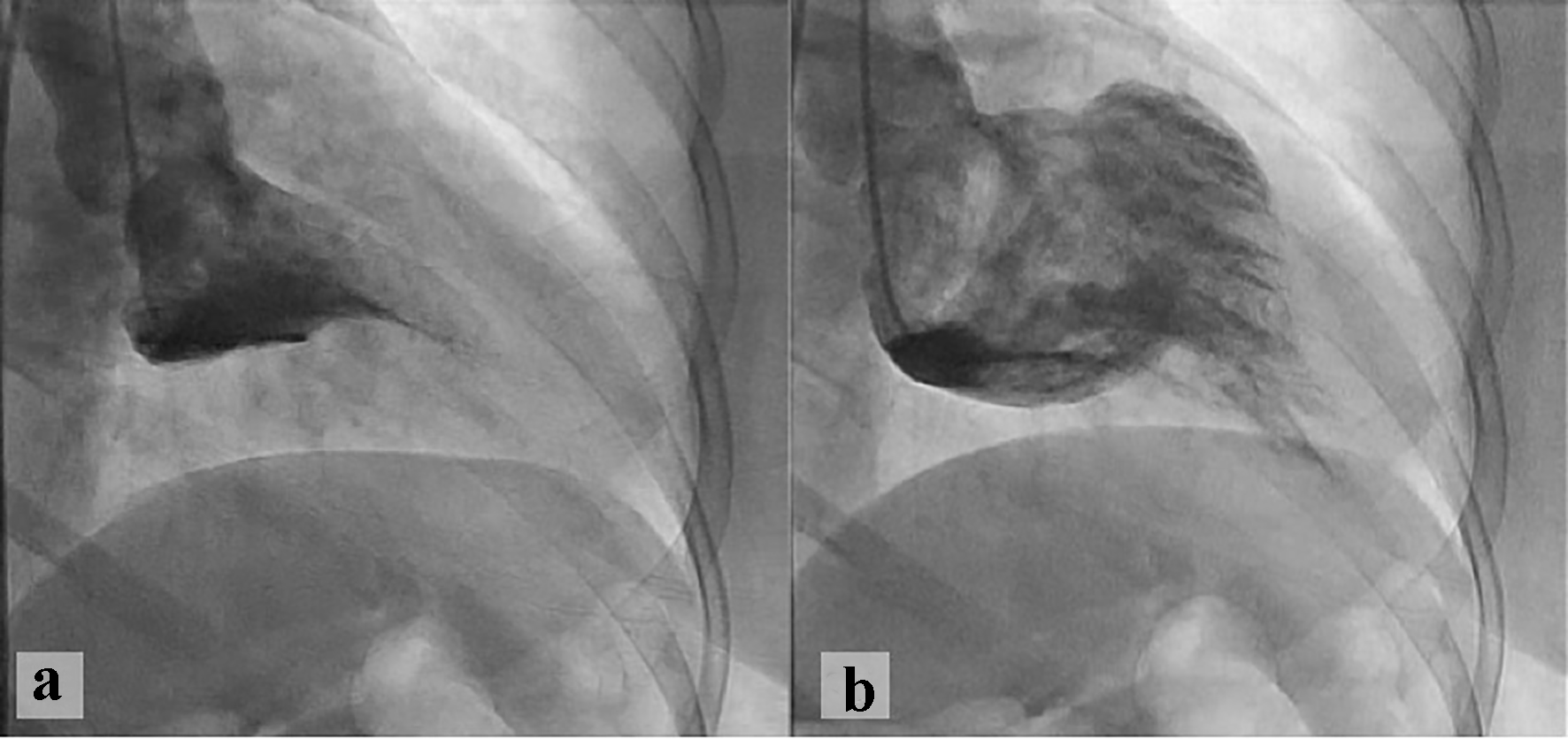
Figures