Figures
![Figure 1.](/tables/cr627w-g001.jpg)
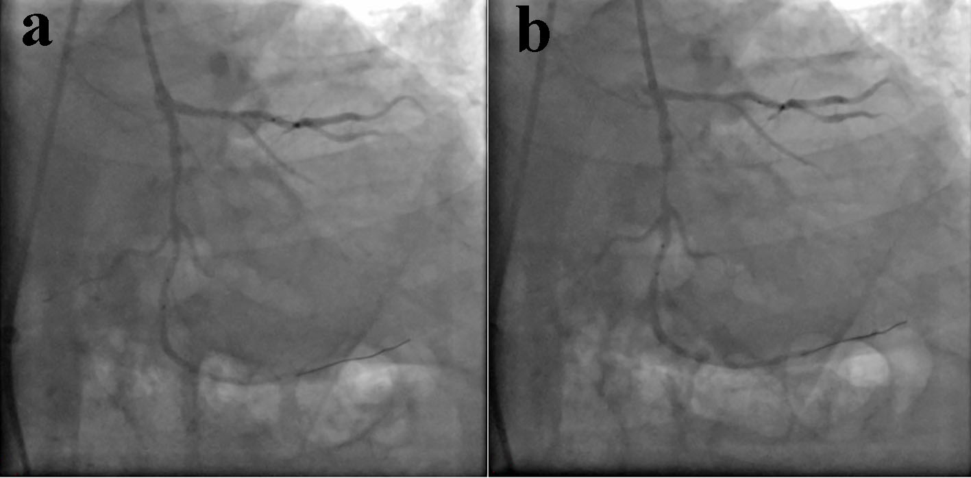
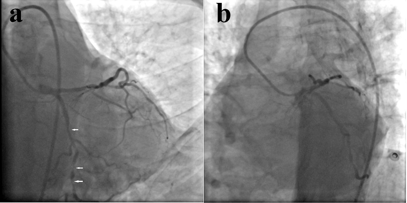
Figure 1. Angiogram showing diffuse, tortuous, calcified narrowing (90% stenosis) in left circumflex (LCx) coronary artery (a: AP caudal view; b: LAO caudal view).
![Figure 2.](/tables/cr627w-g002.jpg)
Figure 2. Lesion was sequentially predilated with multiple balloon at 10 - 15 atm pressure (a, b).
![Figure 3.](/tables/cr627w-g003.jpg)
Figure 3. Morph stent failed to cross the lesion leading to backing of guide catheter (a). Embolized stent visible in right common femoral artery (b).
![Figure 4.](/tables/cr627w-g004.jpg)
Figure 4. Embolized stent further moving down to deep femoral artery (a, b). Judkins right (JR) guiding catheter was carefully parked in right deep femoral artery (b, c).
![Figure 5.](/tables/cr627w-g005.jpg)
Figure 5. Embolized stent being trapped and retrieved (a, b). Whole assembly: stent-snare-guiding catheter was pulled back under fluoroscopic surveillance (c, d).
![Figure 6.](/tables/cr627w-g006.jpg)
Figure 6. Sheath was firmly holded with left hand and whole assembly was successfully pulled out (a, c, c, and d).
![Figure 7.](/tables/cr627w-g007.jpg)
Figure 7. Retrieved stent snared by EN snare (a, b).
![Figure 8.](/tables/cr627w-g008.jpg)
Figure 8. Rewiring of LCx and successful stenting with 3 - 2.5 × 60 mm Morph stent following aggressive predilatation by bigger balloon (a, b, c).