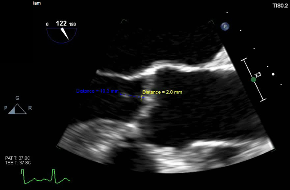
Figure 1. TEE three-chamber view of the mid esophageal region showing 2 mm vegetation along the aortic valve.
| Cardiology Research, ISSN 1923-2829 print, 1923-2837 online, Open Access |
| Article copyright, the authors; Journal compilation copyright, Cardiol Res and Elmer Press Inc |
| Journal website https://www.cardiologyres.org |
Case Report
Volume 9, Number 1, February 2018, pages 59-62
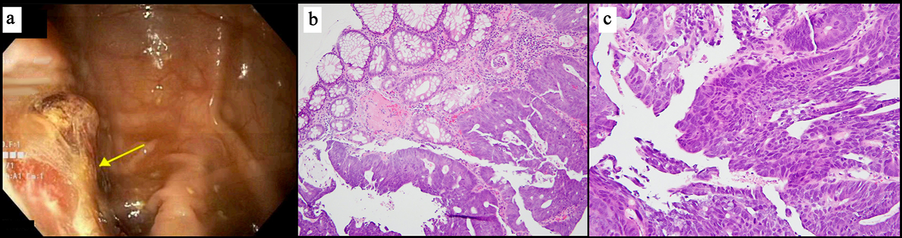
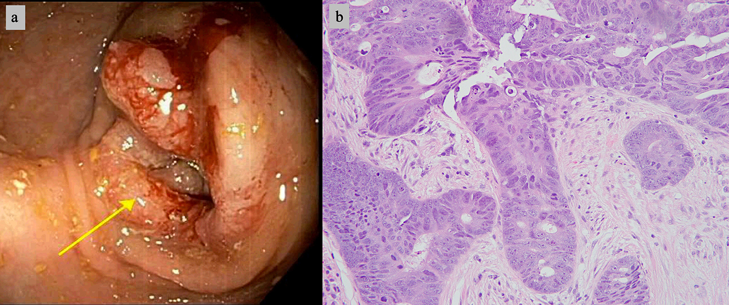
Group G Streptococcus Infective Endocarditis in Association With Colon Cancer
Figures