Cardiology Research, ISSN 1923-2829 print, 1923-2837 online, Open Access
Article copyright, the authors; Journal compilation copyright, Cardiol Res and Elmer Press Inc
Journal website https://www.cardiologyres.org

Case Report

Volume 9, Number 1, February 2018, pages 68-71


Massive Hemorrhagic Pericardial Effusion With Cardiac Tamponade as Initial Manifestation of Mixed Connective Tissue Disease

Figures

Figure 1.
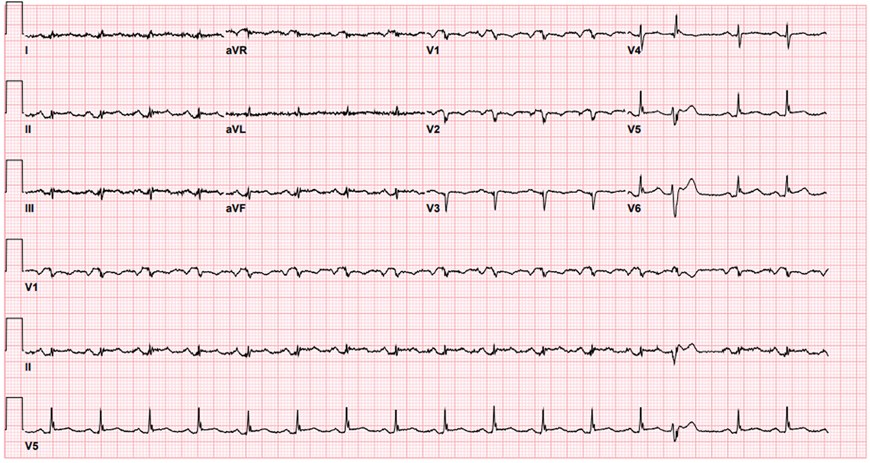
Figure 1. EKG shows sinus rhythm with occasional premature ventricular complexes and low voltage QRS.
Figure 2.
Figure 2. Transthoracic echocardiography, subcostal four chamber view showing diastolic collapse of the RV.
Figure 3.
Figure 3. CT scan of the chest shows pigtail catheter coursing within the pericardium and around the anterolateral heart border with tip terminating posterior to the left atrium. Mild to moderate residual pericardial effusion along the inferior heart border. Bilateral pleural effusions, small to moderate on the left and small on the right. Moderate to severe emphysematous changes.
Figure 4.
Figure 4. Pericardial tissue biopsy showing reactive mesothelial lining correlated with pericarditis.