Cardiology Research, ISSN 1923-2829 print, 1923-2837 online, Open Access
Article copyright, the authors; Journal compilation copyright, Cardiol Res and Elmer Press Inc
Journal website https://www.cardiologyres.org

Original Article

Volume 9, Number 1, February 2018, pages 28-34


The Relationship Between Absence Coronary Artery Calcification and Myocardial Perfusion Single Photon Emission Computed Tomography

Figure

Figure 1.
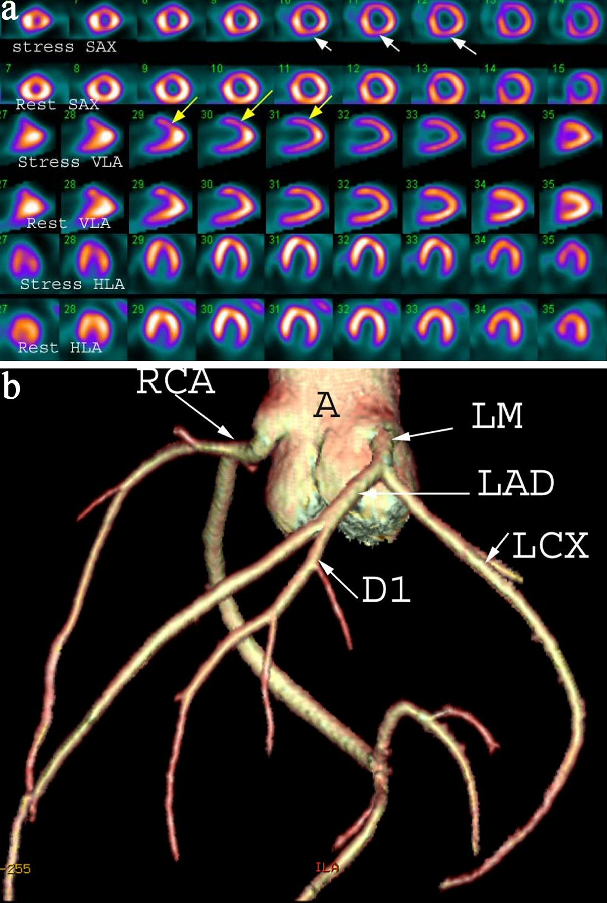
Figure 1. (a) A 42-year-old female with chest pain and shortness of breath. Myocardial perfusion scintigraphy demonstrates reversible defects in the apex and anterior wall (yellow arrows) and inferior wall (white arrows) suggesting inducible ischemia. Gated images (not shown) demonstrate mildly dilated left ventricular cavity and global hypokinesis and impaired left ventricle ejection fraction of 35%. SAX: short axis; VLA: vertical long axis; HLA: horizontal long axis. (b) Shaded surface display coronary computed tomography angiogram demonstrating normal coronary arteries without calcified or non-calcified plaques. A: ascending aorta; LM: left main; LAD: left anterior descending artery; D1: first diagonal artery; LCX: left circumflex artery; RCA: right coronary artery.

Tables

Table 1. Baseline Demographics of The study Population
 
N157
Age (years)53 ± 10
Female88 (56%)
Male69 (44%)
Normal MPS122 (78%)
Abnormal MPS35 (22%)
Diabetes mellitus89 (57%)
Hypertension66 (66%)
Smoking14 (9%)
Hypercholesterolemia33 (33%)
Family history6 (4%)
Left ventricular function60 ± 11%


Table 2. MPS Results
 
N157
Normal MPS122 (78%)
Abnormal MPS35 (22%)
Fixed defect21 (13%)
Equivocal10 (6%)
Reversible defect4 (3%)


Table 3. CT Coronary Angiography Results
 
N35
Normal coronary artery30 (85%)
Coronary artery stenosis
  More than 50%1 (3%)
  Less than 50%1 (3%)
Anomalous coronary artery origin1 (3%)
Myocardial bridging2 (6%)


Table 4. CT Coronary Angiography and Cardiac Morphology Findings in Patients With Abnormal MPS
 
N35
Normal CT coronary angiography and normal cardiac morphology13 (37%)
Abnormal CT coronary angiography and/or abnormal cardiac morphology22 (63%)
Dilated cardiomyopathy14 (40%)
Coronary artery stenosis, more than 50%1 (3%)
Asymmetrical septal hypertrophy1 (3%)
Myocardial bridging2 (6%)
Anomalous coronary artery origin1 (3%)
Mitral valve disease (stenosis, post mitral valve replacement)3 (9%)