Cardiology Research, ISSN 1923-2829 print, 1923-2837 online, Open Access
Article copyright, the authors; Journal compilation copyright, Cardiol Res and Elmer Press Inc
Journal website https://www.cardiologyres.org

Case Report

Volume 9, Number 3, June 2018, pages 176-179


High Grade Atrioventricular Block in Von Recklinghausen Disease: A Rare Phenomenon

Figures

Figure 1.
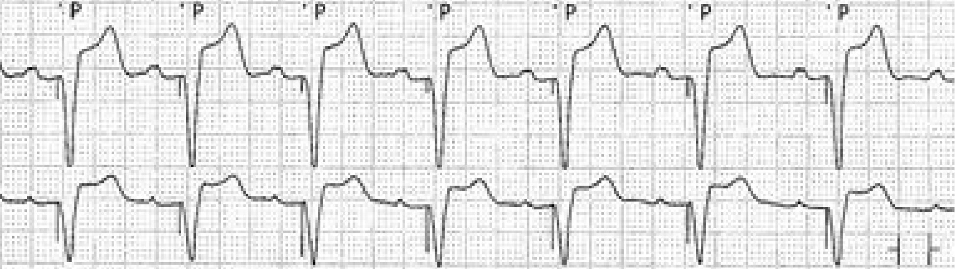
Figure 1. Patient’s twelve-lead electrocardiogram revealing third degree atrioventricular block.
Figure 2.
Figure 2. Parasternal view (left panel) and four chamber view (right panel) transthoracic echocardiogram still images revealing normal left ventricular cavity size with mild concentric hypertrophy. Also, no evidence of cardiac masses was detected.
Figure 3.
Figure 3. Chest radiograph demonstrating subtle mild pulmonary congestion (left panel). Dual chamber pacemaker leads showing insertion in the right atrium and right ventricular trabeculae (right panel).
Figure 4.
Figure 4. Patient’s telemetry strip showing a ventricular paced rhythm after pacemaker insertion.