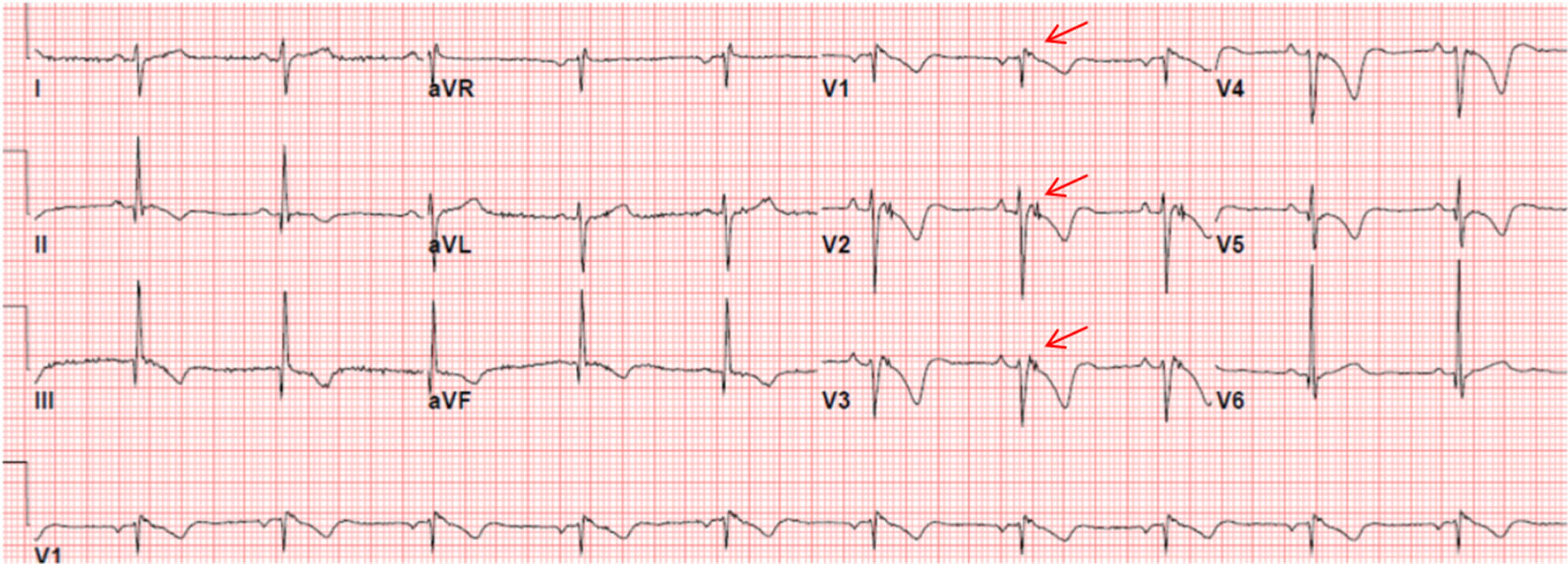
Figure 1. ECG of a patient with ARVC/D showing the presence of an epsilon wave (electric potentials after the end of the QRS complex) (red arrows) and T-wave inversion in V1 - V5.
| Cardiology Research, ISSN 1923-2829 print, 1923-2837 online, Open Access |
| Article copyright, the authors; Journal compilation copyright, Cardiol Res and Elmer Press Inc |
| Journal website https://www.cardiologyres.org |
Original Article
Volume 9, Number 4, August 2018, pages 204-214
Appropriate and Inappropriate Implantable Cardioverter Defibrillators Therapies in Arrhythmogenic Right Ventricular Cardiomyopathy/Dysplasia Patients
Figures
Tables
| Patient characteristics | ICD primary prevention N (%) | ICD secondary prevention N (%) | P value |
|---|---|---|---|
| Male (%) | 6 (85.7 %) | 14 (93.3%) | 0.563 |
| Female (%) | 1 (14.3 %) | 1 (6.7%) | 0.563 |
| Clinical presentation | |||
| Palpitations | 5 (71.4%) | 14 (93.3%) | 0.163 |
| Syncope | 1 (14.3 %) | 2 (13.3%) | 0.952 |
| Shortness of breath | 2 (28.6%) | 2 (13.3%) | 0.388 |
| Chest pain | 1 (14.3 %) | 2 (13.3%) | 0.952 |
| Dizziness | 1 (14.3 %) | 2 (13.3%) | 0.952 |
| Ventricular tachycardia | 0 (0.0%) | 12 (80%) | 0.000 |
| Sudden cardiac death | 0 (0.0%) | 3 (20.0%) | 0.203 |
| Medications | |||
| Amiodarone | 0 (0.0%) | 3 (20.0%) | 0.203 |
| Sotalol | 1 (14.0%) | 6 (40.0%) | 0.228 |
| Beta blockers | 4 (57.1%) | 7 (46.7%) | 0.647 |
| Angiotensin-converting enzyme inhibitors (ACE inhibitors) | 2 (28.6%) | 7 (46.7%) | 0.421 |
| Spironolactone | 0 (0.0%) | 2 (13.3%) | 0.311 |
| Implantable cardioverter defibrillator (ICD) therapy | |||
| Single chamber ICD | 3 (42.9%) | 10 (66.7%) | 0.296 |
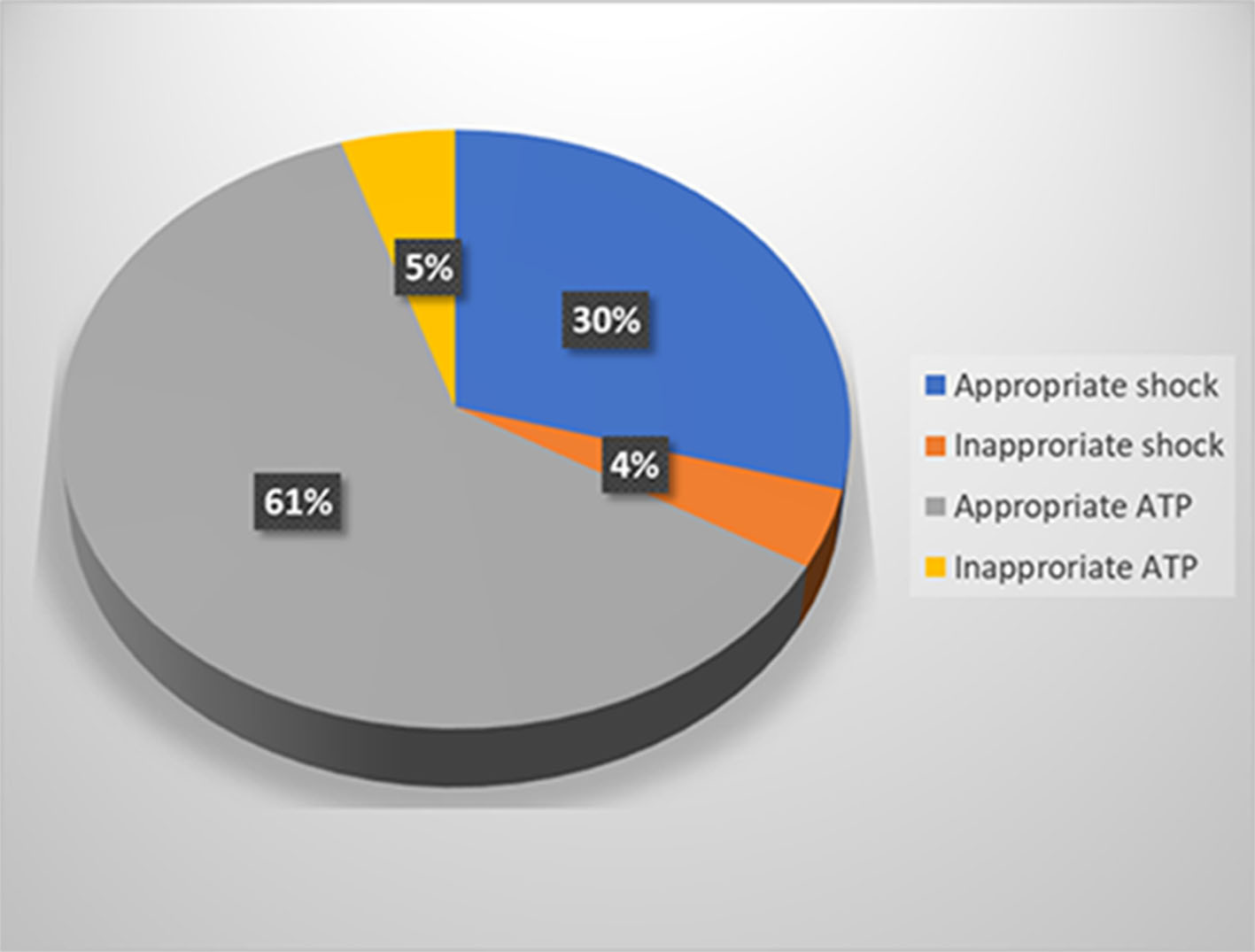
| Appropriate ICD therapy | 1 (14.0%) | 10 (66.7%) | 0.022 |
| Inappropriate ICD therapy | 1 (14.0%) | 4 (26.7%) | 0.519 |
| Genetic results | |||
| Positive | 3 (13.6%) | 7 (31.8%) | 0.672 |
| Negative | 2 (9.1%) | 2 (9.1%) | |
| Not performed | 2 (9.1%) | 6 (27.3%) | |
| Patients | Epsilon wave (M)* | T-wave inversion in V1-3 or beyond (M)* | T-wave inversion in V1-2 or V4, 5 or 6 (m)* | RBBB + T-wave inversion in V1-4 (m)* | Terminal activation duration of QRS in V1, 2 or 3 ≥ 55 ms (m)* |
|---|---|---|---|---|---|
| *M indicates major, and m indicates minor diagnostic criterion based on 2010 ARVC/D Task Force Criteria [4]. RBBB: right bundle-branch block. -: absent; +: present. | |||||
| 1 | + | + | - | - | + |
| 2 | - | + | - | - | - |
| 3 | - | + | - | - | - |
| 4 | - | + | - | - | - |
| 5 | + | + | - | - | - |
| 6 | - | + | - | - | - |
| 7 | - | + | - | - | - |
| 8 | + | + | - | - | - |
| 9 | - | - | - | - | - |
| 10 | + | + | - | - | - |
| 11 | - | + | - | - | - |
| 12 | - | - | - | - | - |
| 13 | - | - | + | - | + |
| 14 | - | - | - | - | - |
| 15 | + | + | - | - | + |
| 16 | - | - | + | - | + |
| 17 | - | + | - | - | - |
| 18 | - | + | - | - | + |
| 19 | + | + | - | - | - |
| 20 | - | + | - | - | - |
| 21 | - | - | - | - | - |
| 22 | - | - | - | - | + |
| Patients no. | LVEF | RV systolic function | PLAX RVOT (corrected for body size (PLAX/BSA)) | PSAX RVOT (corrected for body size (PSAX/BSA)) | Other abnormalities |
|---|---|---|---|---|---|
| BSA: body surface area; LVEF: Left ventricular ejection fraction; mm: millimeters; m2: square meter; PLAX: parasternal long-axis view; PSAX: parasternal short-axis view; PR: pulmonic valvular regurgitation; RV: right ventricle; RVOT: RV outflow tract; SPAP: systolic pulmonary artery pressure; TR: tricuspid regurgitation. For the 2D echo major and minor criteria values, see reference [4]. | |||||
| 1 | > 55% | Mildly reduced | 43 mm (21.5 mm/m2) | 46 mm (23 mm/m2) | |
| 2 | 45-50% (47.5%) | Moderately to severely reduced | 51 mm (24.3 m/m2) | 48 mm (29 mm/m2) | RV apical trabeculations and small aneurysms |
| 3 | 50-55% (52.5%) | Mildly to moderately reduced | 33 mm (17.3 mm/m2) | 32 mm (16.8 mm/m2) | RV wall thinning, multiple microaneurysms and hyertrabeculations. Mild TR |
| 4 | > 55% | Moderately reduced | 34 mm (16 mm/m2) | 41 mm (19 mm/m2) | RV thinned and akinetic wall, multiple microaneurysms and prominent trabeculations Moderate TR |
| 5 | > 55% | Mildly reduced | 42 mm (23.3 mm/m2) | 45 mm (25 mm/m2) | RV regional hypokinesia Mild TR |
| 6 | > 50% | Severely reduced | 34 (22.7 mm/m2) | 36 mm (24 mm/m2) | Severe TR |
| 7 | > 55% | Mildly reduced | 28 mm (14 mm/m2) | 31 mm (15.5 mm/m2) | RV thin wall with multiple microaneurysms in the apex and RVOT prominent, disarrayed trabeculations and dyskinetic anterior wall of RVOT |
| 8 | 50-55% | Moderately to severely reduced | 30 mm (15.8 mm/m2) | 35 mm (18.4 mm/m2) | Thin and fibrosed RV wall with prominent trabeculations Moderate TR SPAP 35 - 40 mm Hg. |
| 9 | > 55% | Normal | 20 mm (10 mm/m2) | 24 mm (12 mm/m2) | |
| 10 | 30-35% | Severely reduced | 41 mm (34.2 mm/m2) | 40 mm (33.3 mm/m2) | Hypertrabeculated RV Severe TR Mild-to-moderate PR |
| 11 | 45 % | Severely reduced | 33 mm (19.4 mm/m2) | 33 mm (19.4 mm/m2) | Thinning of the RV free wall |
| 12 | > 55% | Normal | 30 mm (18.7 mm/m2) | 34 mm (21.25 mm/m2) | |
| 13 | > 55% | Low normal | 29 mm (15.26 mm/m2) | 28 mm (14.7 mm/m2) | Thinning of the apical RV wall |
| 14 | 51% | Normal | 23 mm (11.5 mm/m2) | 30 mm (15 mm/m2) | RV systolic pressure 30 - 40 mm Hg |
| 15 | 44% | Normal | 32 mm (20 mm/m2) | 39 mm (24.4 mm/m2) | |
| 16 | 45% | Normal | 29 mm (16 mm/m2) | 31 mm (17 mm/m2) | RV systolic pressure 30- 35 mmHg |
| 17 | > 55% | Normal | 28 mm (18.7 mm/m2) | 30 mm (20 mm/m2) | |
| 18 | > 55% | Moderately to severely reduced | 36 mm (24 mm/m2) | 36 mm (24 mm/m2) | Mild TR |
| 19 | > 55% | Mildly reduced | 23 mm (13.5 mm/m2) | 28 mm (16.5 mm/m2) | |
| 20 | < 25% | Severely reduced | 47 mm (29.4 mm/m2) | 48 mm (30 mm/m2) | Markedly trabeculated RV |
| 21 | > 55% | Normal | 27 mm (14 mm/m2) | 24 mm (12.6 mm/m2) | |
| 22 | > 55% | Normal | 21 mm (17.5 mm/m2) | 15 mm (12.5 mm/m2) | |
| Study no./gender | Left ventricle (LV) | Right ventricle (RV) |
|---|---|---|
| *Between practices values corrected for body size (value/body surface area (BSA)). EF: ejection fraction; EDV: end-diastolic volume; ESV: end-systolic volume mm millimeters; m2: square meter, TR: tricuspid valve. For the cardiac MRI major and minor criteria values, see reference [4]. | ||
| 1/M | Normal with EF 55% | EDV 151 mL (79.5 mL/m2)* ESV 83 mL (43.7 mL/m2) RVEF 45% Moderately to severely dilated with dyskinetic motion in the free wall and basal aneurysm T1-weighted images show area of severe thinning on the free wall of right ventricle |
| 2/M | Normal EDV 153 mL ESV 56 mL EF 63% | EDV 178 mL (93.7 mL/m2) ESV 121 mL (63.7 mL/m2) EF 32% Moderate dilatation with generalized hypokinesis. There are localized RV areas of micro-aneurysms, mainly at the RVOT and the RV apex Prominent papillary muscle and the trabeculation |
| 3/M | EF > 55% | EDV 100 mL (50 mL/m2) ESV 48 mL (24 mL/m2) EF 52% Thinned of the free wall with a small area of severe thinning, nears the RV apex with dyskinesis and diastolic bulging fulfilling the criterion of aneurysm |
| 4/F | Normal systolic function | EDV 249 mL (207 mL/m2) ESV 212 mL (176.7 mL/m2) EF 15% Stroke volume 36.2 mL Severely dilated and hypertrabiculated Very thinned out RV especially apical regions with evidence of microaneurysm and diastolic bulge and segmental dilatation of RV The RVOT is dilated with hypokinesia. Evidence of at least mild-to-moderate TR regurgitation |
| 5/F | EDV 83 mL ESV 41 mL EF 50% Mild global hypokinesis and akinesis of the distal septum | EDV 120 mL (70.6 mL/m2) RV ESV 83 mL (48.8 mL/m2) EF 31% Stroke volume 37 mL Severely dilated with severe global hypokinesis and dyskinesis of the RV apex and severe thinning noted in both cine and dark blood imaging Delayed myocardial enhancement demonstrates severe thinning of the free wall of the right ventricle |
| 6/M | EDV 99 mL ESV 39 mL EF 60% Stroke volume 59 mL | EDV 81 mL (50.6 mL/m2) ESV 52 mL (32.4 mL/m2) RVEF 36% Mildly-to-moderately dilated with dyskinesia and akinesis of the free wall adjacent to the right ventricular apex T1, T2 and delayed gadolinium enhancement demonstrate two focal aneurysms in the free wall of RV |
| 7/M | Normal EF 58% | EDV 171 mL (90 mL/m2) ESV 93 mL (49 mL/m2) EF 49% Stroke volume 76 mL( 40 mL/m2) Focal dyskinesia in the RVOT Small patent ductus arteriosus measuring about 5 mm. Main pulmonary artery (MPA) not enlarged measuring 26 mm |
| 8/M | Normal EF 57% | EDV 87 mL (43.5 mL/m2) ESV 47 mL (23.5 mL/m2) EF 46% Mildly dilated with mild global hypokinesis Focal aneurysm noted in RV free wall or RVOT |
| 9/M | Normal LV | Moderately dilated RV and reduced RV function (CMRI was performed in another cardiac center and full report no available) |
| 10/M | EDV 110.5 mL (65 mL/m2) ESV 49.3 mL (29 mL/m2) EF 54% | EDV187.5 mL (105 mL/m2) ESV 100 mL(59 mL/m2) EF 44% |
| 11/M | EF 45-50% | Quantification was not apparent at this point due to the poor ECG gating Dilated at the RVOT, there is evidence of macroaneurysm with small microaneurysm noted at the RV apex with a large macroaneurysm noted at the RVOT and RV free wall with dyskinesis Overall RV systolic function appeared to be mildly reduced |
| 12/M | EDV 80 mL ESV 25 mL EF 68% | EDV 74 mL ESV 26 mL EF 64% Late gadolinium enhancement in mid myocardium at the septum |
| Patient characteristics | Appropriate therapy N (%), 11 (50%) | Inappropriate therapy N (%), 3 (13.6%) | P value |
|---|---|---|---|
| ACE: angiotensin-converting enzyme; ICD: implantable cardioverter defibrillator. | |||
| Male (%) | 10 (90.9 %) | 3 (100%) | 0.814 |
| Female (%) | 1 (9.1 %) | 0 (0.0%) | 0.814 |
| Clinical presentation | |||
| Palpitations | 10 (90.9 %) | 3 (100%) | 0.462 |
| Syncope | 1 (9.1 %) | 1 (33.3%) | 0.552 |
| Shortness of breath | 2 (18.2%) | 1 (33.3%) | 0.727 |
| Chest pain | 2 (18.2%) | 1 (33.3%) | 0.295 |
| Dizziness | 1(9.1 %) | 1(33.3%) | 0.552 |
| Ventricular tachycardia | 8 (72.7 %) | 1 (33.3%) | 0.229 |
| Sudden cardiac death | 3(27.3%) | 0 (0.0%) | 0.176 |
| Medications | |||
| Amiodarone | 2 (18.2%) | 0 (0.0%) | 0.713 |
| Sotalol | 2 (18.2%) | 0 (0.0%) | 0.713 |
| Beta blockers | 4 (36.4%) | 1 (33.3%) | 0.870 |
| ACE inhibitors | 6 (54.5%) | 2 (66.7%) | 0.630 |
| Spironolactone | 6 (54.5%) | 1 (33.3%) | 0.416 |
| ICD type | |||
| Single ICD | 2 (18.2%) | 0 (0.0%) | 0.333 |
| Dual ICD | 9 (81.8%) | 3 (100%) | |
| Genetic results | |||
| Positive | 6 (54.5%) | 2 (66.7%) | 0.323 |
| Negative | 1 (9.1 %) | 1 (33.3%) | 0.516 |
| Not performed | 4 (36.4%) | 0 (0.0%) | 0.308 |
| Prevention type | |||
| Primary | 1 (9.1 %) | 1 (33.3%) | 0.048 |
| Secondary | 10 (90.9%) | 2 (66.7%) | |