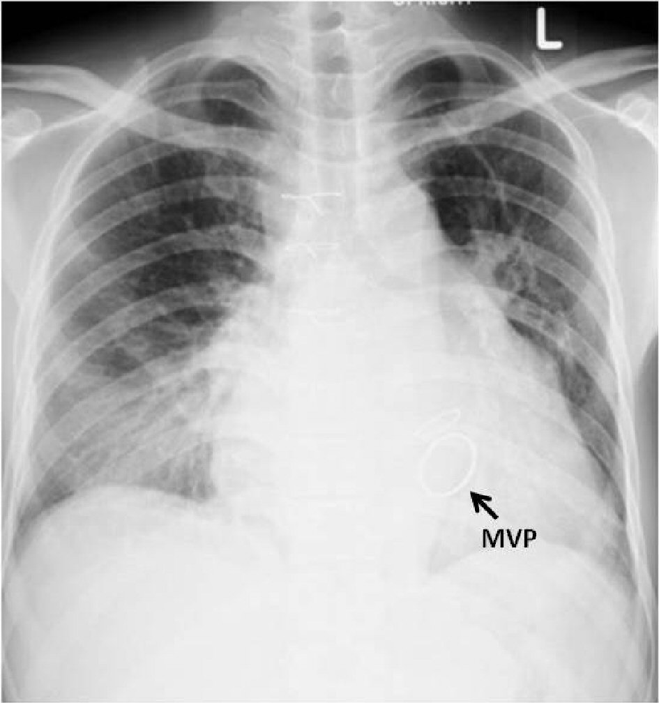
Figure 1. Chest X-ray showing cardiomegaly with pulmonary congestion and mitral valve prosthesis (MVP).
| Cardiology Research, ISSN 1923-2829 print, 1923-2837 online, Open Access |
| Article copyright, the authors; Journal compilation copyright, Cardiol Res and Elmer Press Inc |
| Journal website https://www.cardiologyres.org |
Case Report
Volume 9, Number 5, October 2018, pages 314-317
Corynebacterium diphtheriae Native Aortic Valve Endocarditis in a Patient With Prosthetic Mitral Valve: A Rare Presentation
Figures
Table
| Results | |
|---|---|
| CBC: complete blood count; HCT: hematocrit; WBC: white blood cell; LFT: liver function test; AST: aspartate aminotransferase; ALT: alanine aminotransferase. | |
| CBC | Hb: 12.8 g/dL, HCT: 34.8%, WBC: 8,800 / µL; N: 86%, L: 10%, Mono: 2%, band form: 2%, platelets: 153,000/µL |
| Kidney function | BUN: 12.4 mg/dL, creatinine: 1.0 mg/dL |
| LFT | Bilirubin ( total): 4.3 µmol/L, bilirubin (direct): 3.7 µmol/L, AST: 41.0 IU/L, ALT: 18 IU/L, AP; 230 IU/L, protein: 5.6 mg/dL, albumin: 2.8 mg/dL |
| Electrolyte | Na: 128.0 mmol/L, K: 4.0 mmol/L, Cl: 86 mmol/L, HCO3 29 mEq/L |