Cardiology Research, ISSN 1923-2829 print, 1923-2837 online, Open Access
Article copyright, the authors; Journal compilation copyright, Cardiol Res and Elmer Press Inc
Journal website http://www.cardiologyres.org

Case Report

Volume 9, Number 6, December 2018, pages 381-384


Pericarditis After Breast Implant Rupture: A Case Report

Figures

Figure 1.
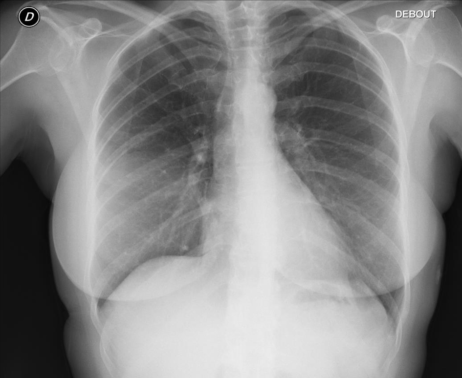
Figure 1. Chest X-ray showing no abnormalities but an asymmetry between the right and left breast.
Figure 2.
Figure 2. Echocardiography showing an apical pericardial effusion in apical four-chamber view (a, red stars) and subcostal view (b, red arrow).
Figure 3.
Figure 3. MRI showing the rupture of the implant in the left breast. (a) T2 propeller 3-0 sequence allowing minimizing the motion artifacts and showing a silicone granuloma. (b) Axial sequence T2 short T1 inversion recovery allowing the suppression of the fat signal. (c) Axial sequence T1. b and c show an effusion inside the silicone of the implant, indicating the rupture of both the implant and the periprosthetic capsule.