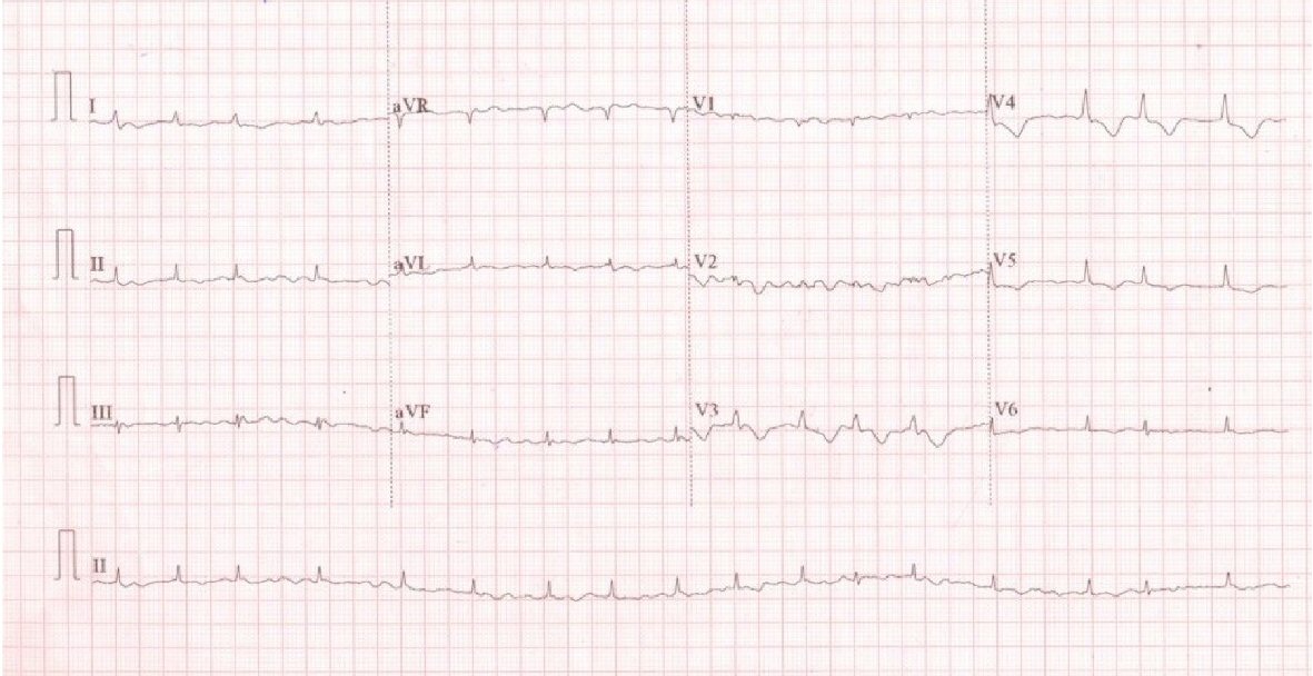
Figure 1. ECG showed atrial fibrillation and controlled ventricular rate.
| Cardiology Research, ISSN 1923-2829 print, 1923-2837 online, Open Access |
| Article copyright, the authors; Journal compilation copyright, Cardiol Res and Elmer Press Inc |
| Journal website http://www.cardiologyres.org |
Case Report
Volume 10, Number 2, April 2019, pages 124-127
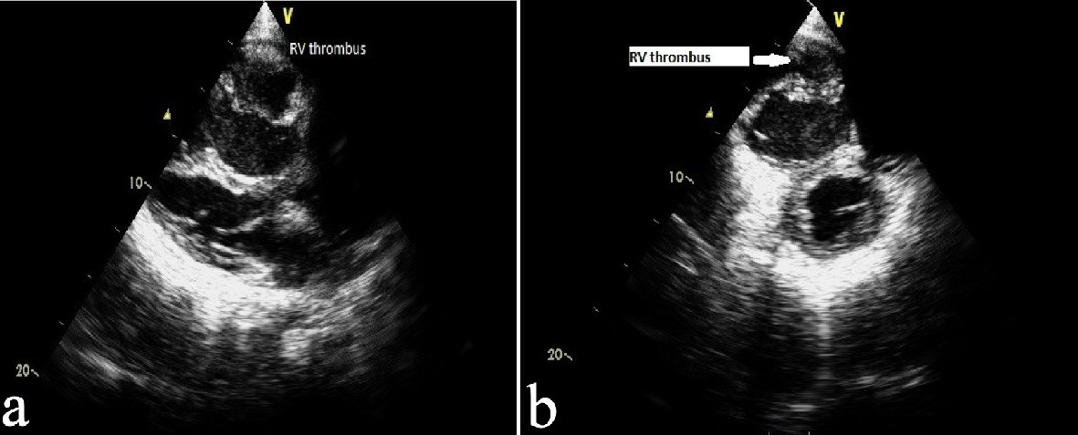
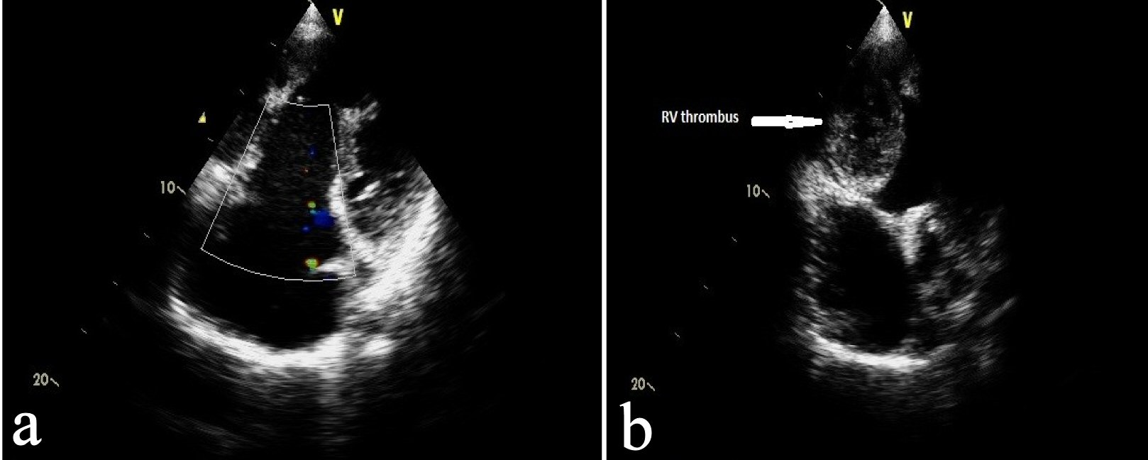
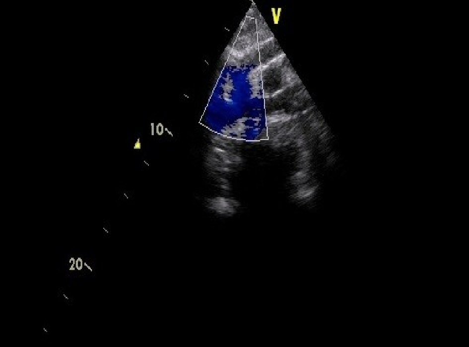
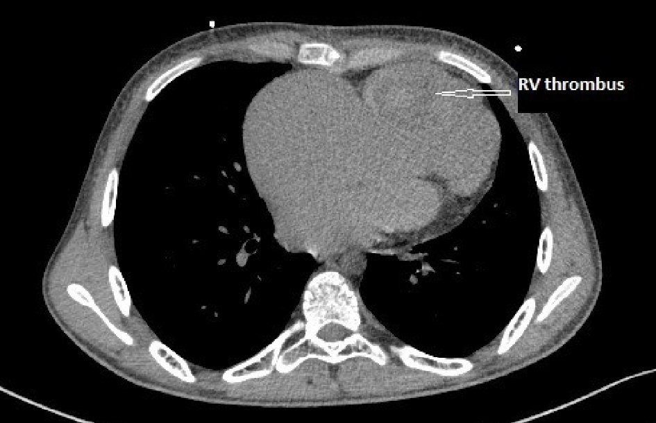
Large Right Ventricle Thrombus in Uhl’s Anomaly: A Rare Presentation of Extremely Rare Disease
Figures