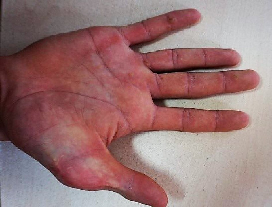
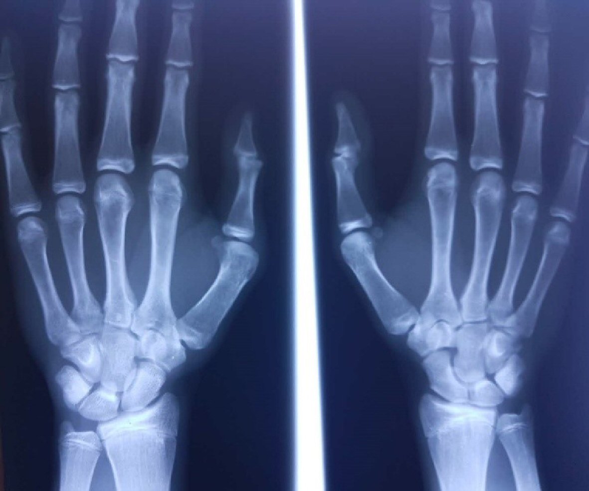
Figure 1. Right hand: surgical scar of the thumb.
| Cardiology Research, ISSN 1923-2829 print, 1923-2837 online, Open Access |
| Article copyright, the authors; Journal compilation copyright, Cardiol Res and Elmer Press Inc |
| Journal website https://www.cardiologyres.org |
Case Report
Volume 9, Number 5, October 2018, pages 324-329
Holt-Oram Syndrome With Multiple Cardiac Abnormalities
Figures
Table
| Main clinical characteristics | Defects | Observations |
|---|---|---|
| Upper-limb malformation | Carpal bones malformations | 100% |
| Triphalangeal or absent thumb(s) | ||
| Preaxial polydactyly (duplication of the thumb) | ||
| Aplasia/hypoplasia of the radius | ||
| Abnormal forearm pronation and supination | ||
| Abnormal opposition of the thumb | ||
| Sloping shoulders/restriction of shoulder joint movement | ||
| Congenital heart defect | ASD | 75% ASD: the most common |
| VSD | ||
| Pulmonary atresia/stenosis | ||
| Double outlet right ventricle | ||
| Aortic valve insufficiency | ||
| Aortic valve stenosis | ||
| Tricuspid atresia | ||
| Mitral valve abnormality | ||
| Patent ductus arteriosus | ||
| Pentalogy of Fallot | ||
| Tetralogy of Fallot | ||
| Common arterial truncus | ||
| Dextrocardia | ||
| Right aortic arch | ||
| Cardiac conduction disease | Sinus bradycardia | |
| Atrioventricular (AV) block | ||
| Atrial fibrillation |