Cardiology Research, ISSN 1923-2829 print, 1923-2837 online, Open Access
Article copyright, the authors; Journal compilation copyright, Cardiol Res and Elmer Press Inc
Journal website http://www.cardiologyres.org

Case Report

Volume 9, Number 6, December 2018, pages 395-399


A Successfully Treated STEMI Due to Simultaneous Thrombotic Occlusion of Left Anterior Descending Artery and Left Circumflex Artery: A Case Report and Review of the Literature

Figures

Figure 1.
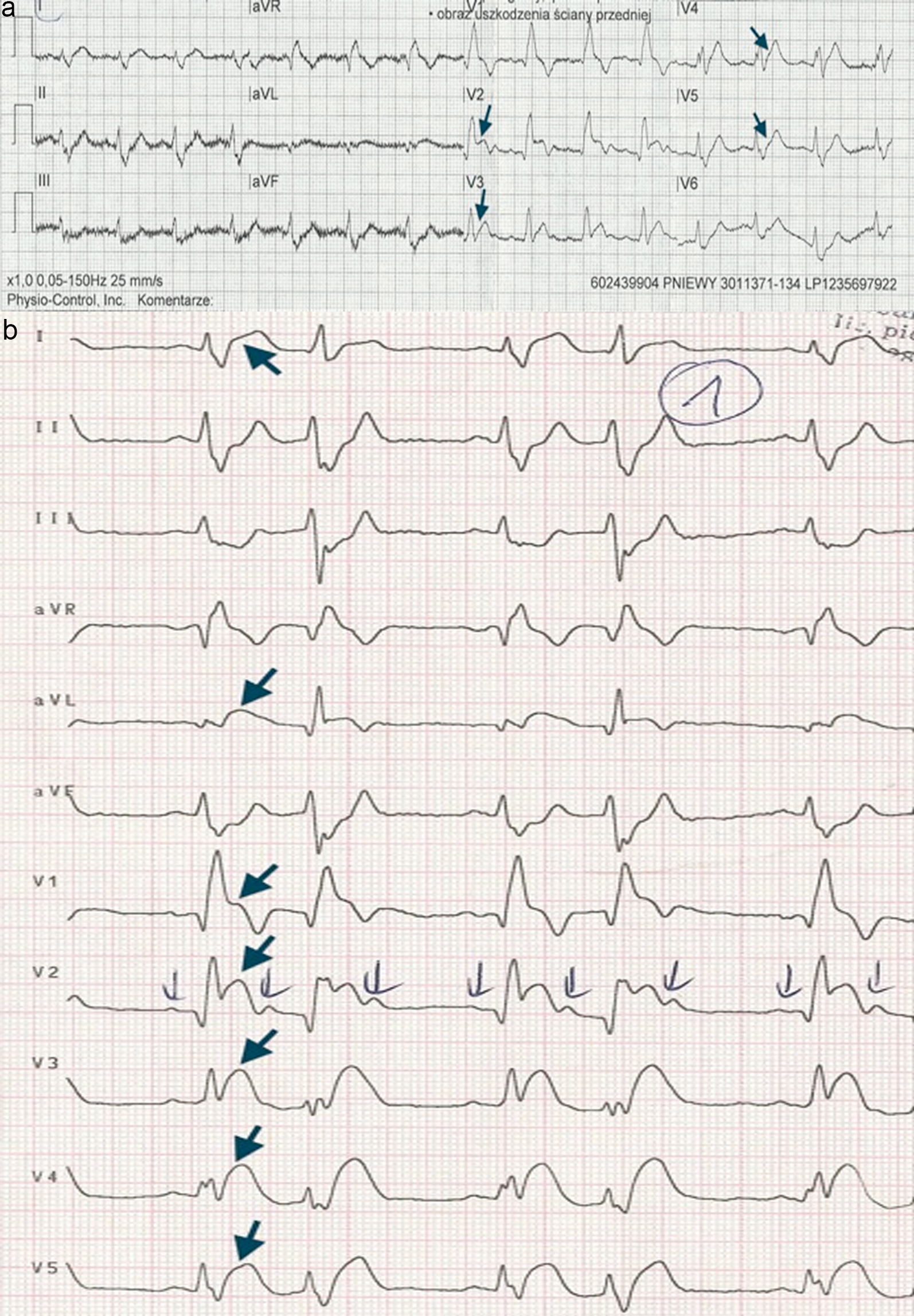
Figure 1. The patient’s ECG. (a) ECG transmitted to the cath lab using LifeNet system (Physio Control, USA). ST-segment elevation in precordial leads V2 - V5 (black arrows). (b) ECG obtained immediately after arrival to the hospital. ST-segment elevation in precordial leads V1 - V6 and leads I, aVL (black arrows).
Figure 2.
Figure 2. Coronary angiography of left coronary artery - a simultaneous occlusion of LAD (white arrow) and LCx (black arrow), a critical stenosis of first marginal branch (black arrow with interrupted line). LAD: left anterior descending artery; LCx: left circumflex artery.
Figure 3.
Figure 3. Coronary angiography of RCA - a significant stenosis in its mid-portion (white arrow). RCA: right coronary artery.
Figure 4.
Figure 4. Coronary angiography of left coronary artery following PCIs of both infarct-related arteries (LAD and LCx). PCIs: percutanous coronary interventions; LAD: left anterior descending artery; LCx: left circumflex artery.