Figures
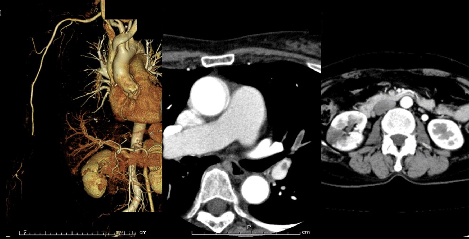
Figure 1. Enhanced computed tomography of the patient. 3D-CT scan shows interruption of the blood flow in the distal part of brachial artery and development of collaterals circulation (left photo). Contrast CT scan shows left pulmonary artery thromboembolism (middle photo) and right kidney infraction (right photo). CT: computed tomography.
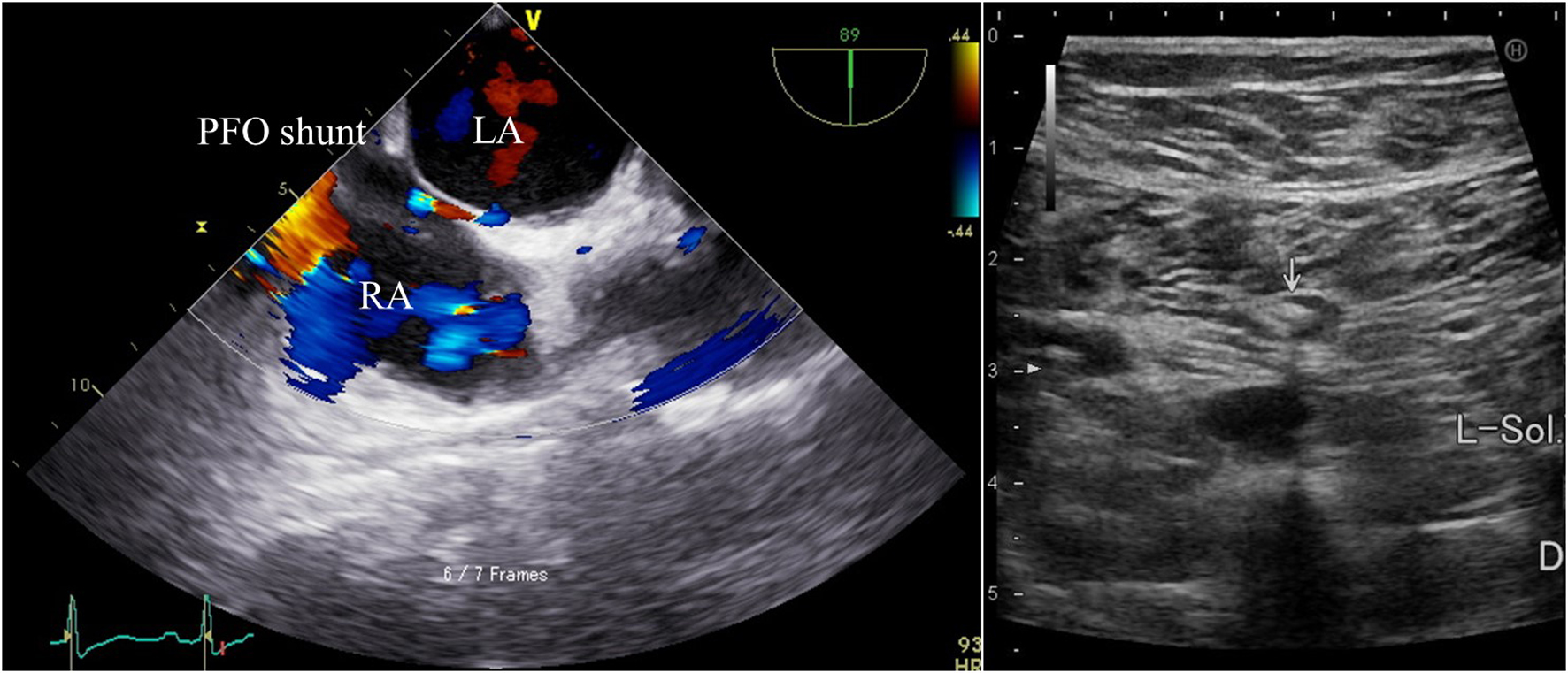
Figure 2. The imaging of transesophageal echocardiography and venous ultrasonography. Transesophageal echocardiography (left photo) shows the presence of patent foramen ovale (PFO) and right to left shunt in Valsalva maneuver. Interatrial septal aneurysm and left atrial thrombus are not observed. Venous ultrasonography (right photo) shows presence of deep vein thrombosis in left soleal vein. LA: left atrium; RA: right atrium.
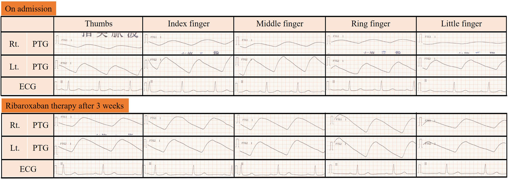
Figure 3. The imaging of photoplethysmogram. The figure shows photoplethysmogram (PTG) of patient fingers. Upper section is her right hand, lower section is her left hand, finger number from right to left. On admission, her PTG was slow. After ribaroxaban therapy for 3 weeks, her PTG was normalized. PTG: photoplethysmogram; ECG: electrocardiogram.
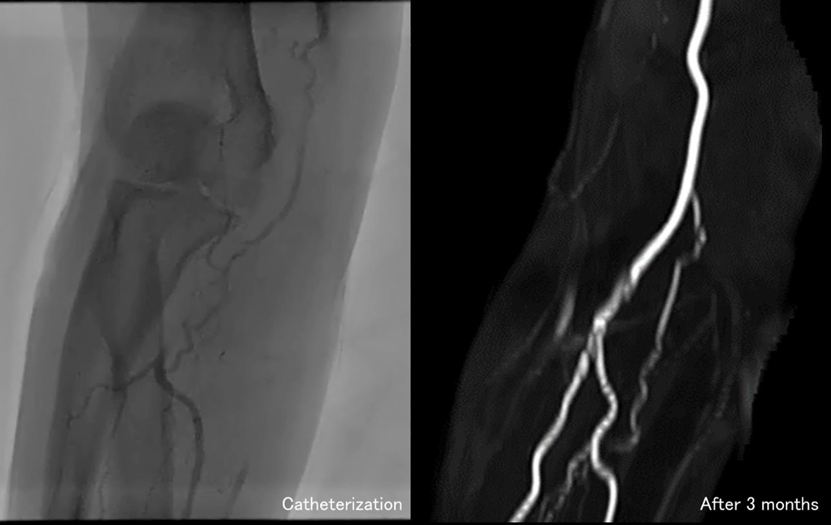
Figure 4. The imaging of brachial artery angiography. Catheterization shows the thrombus still remains in brachial artery and development of collateral circulation on the seventh day after admission (left photo). Three months after discharge, MRA shows brachial artery could achieve reperfusion (right photo). MRA: magnetic resonance angiography.
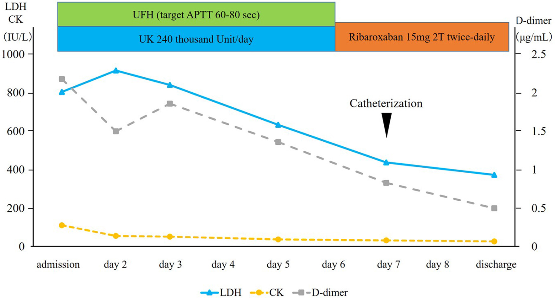
Figure 5. Clinical course of the patient. UFH: unfractionated heparin; UK: urokinase; APTT: activated partial thromboplastin time; LDH: lactate dehydrogenase; CK: creatine kinase.