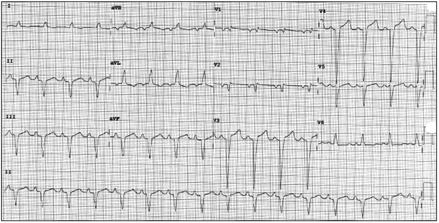
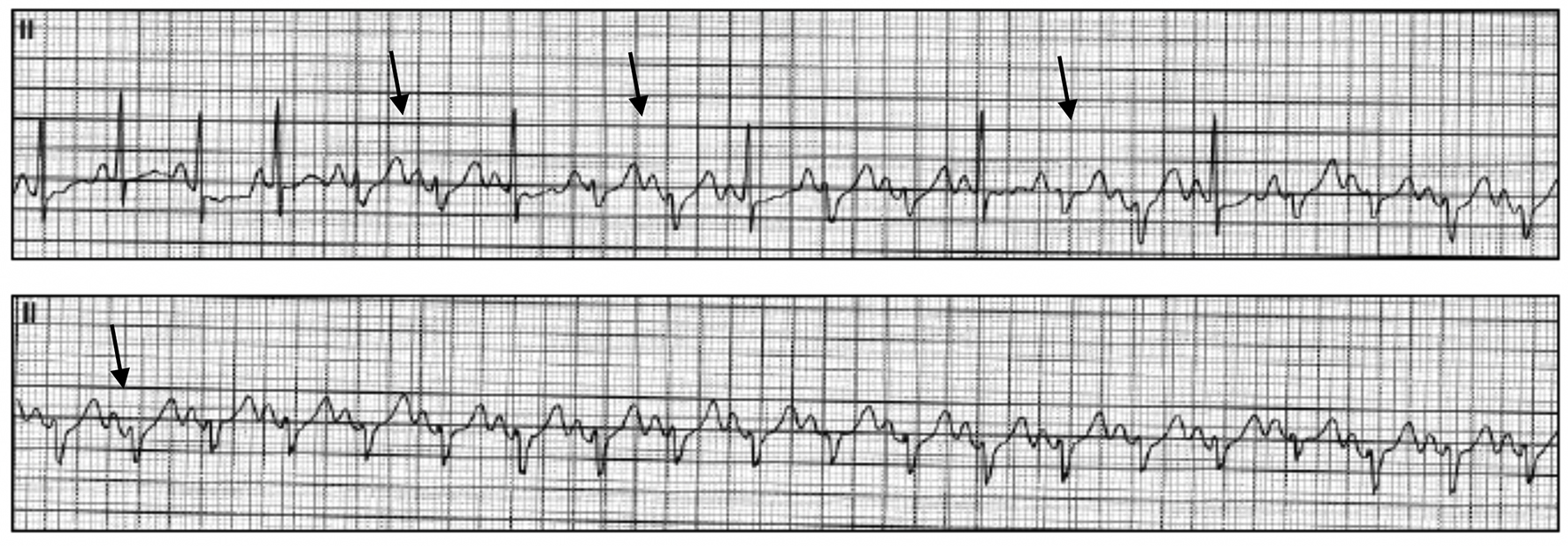
Figure 1. Presenting ECG to cardiology clinic with NSR and newly recognized LBBB. ECG: electrocardiogram; NSR: normal sinus rhythm; LBBB: left bundle branch block.
| Cardiology Research, ISSN 1923-2829 print, 1923-2837 online, Open Access |
| Article copyright, the authors; Journal compilation copyright, Cardiol Res and Elmer Press Inc |
| Journal website http://www.cardiologyres.org |
Case Report
Volume 10, Number 4, August 2019, pages 230-235
Trastuzumab-Induced Cardiomyopathy and Intermittent Left Bundle Branch Block
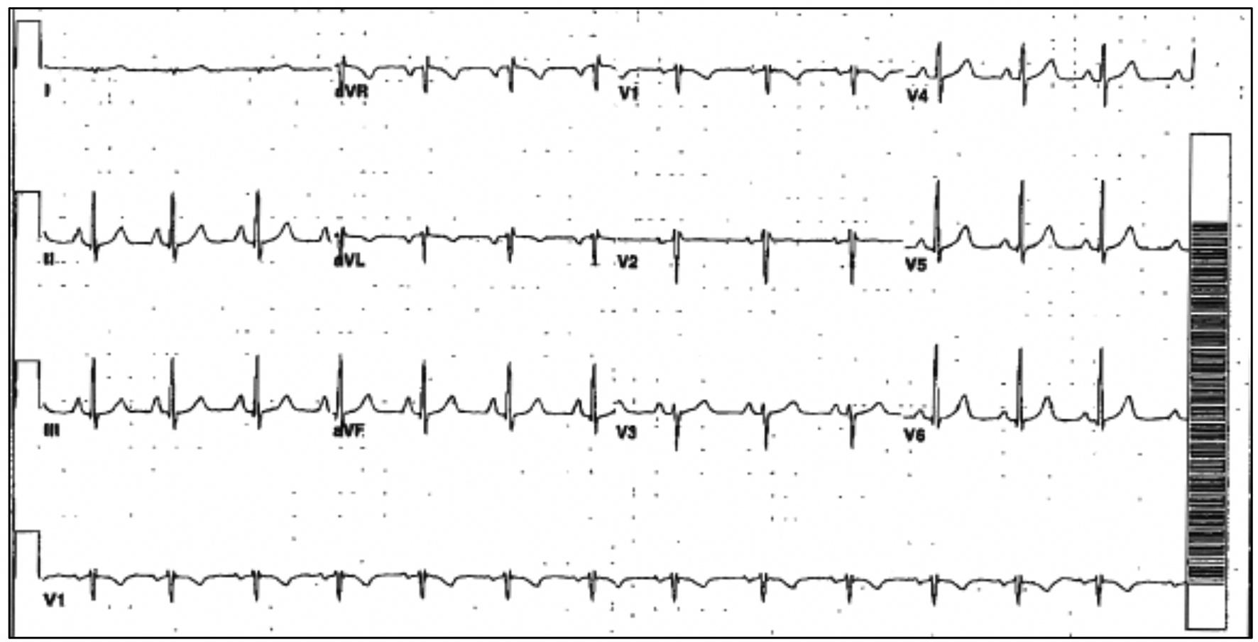
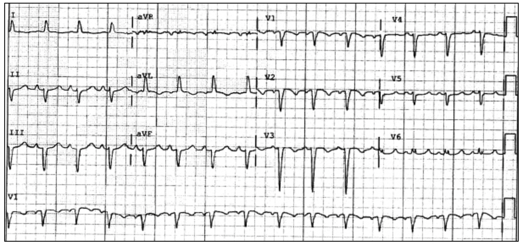
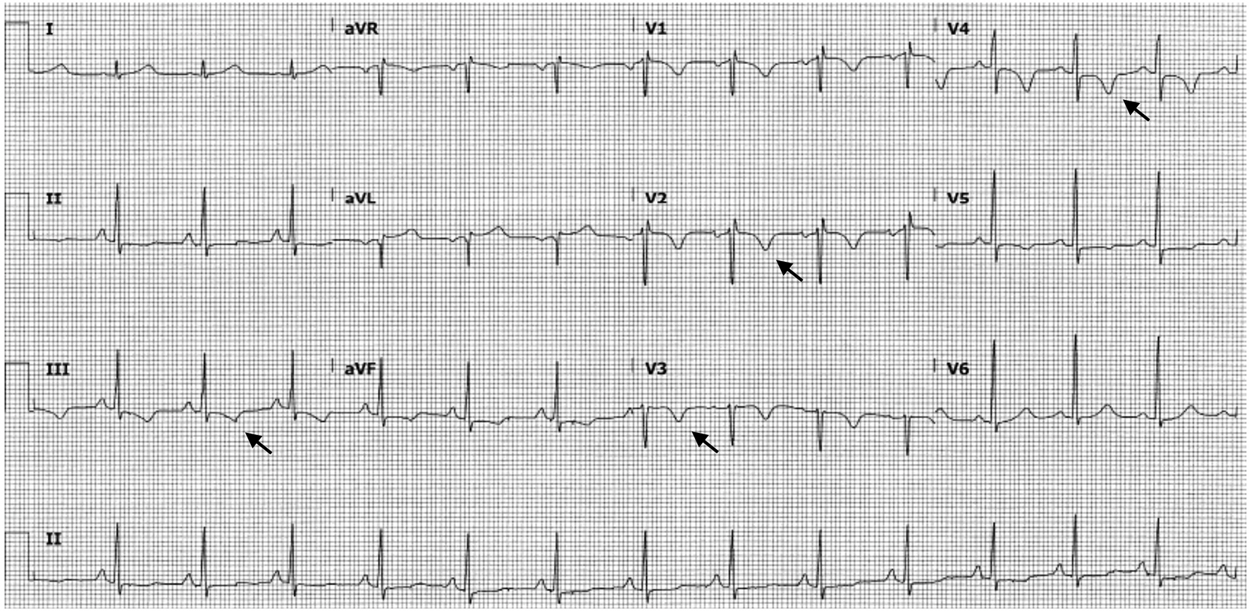
Figures
Table
| Complications | Study | Symptoms | Further testing and results | Cardiomyopathy present |
|---|---|---|---|---|
| LBBB: left bundle branch block; RBBB: right bundle branch block; LV: left ventricular; NSVT: non-sustained ventricular tachycardia. | ||||
| Asymptomatic new LBBB | Piotrowski et al [6] | No symptoms | No | No |
| New RBBB | Piotrowski et al [6] | No Symptoms | No | No |
| T-wave inversions in anterior leads | Olin et al [13] | Chest pain | Left heart catheterization revealed normal coronaries. | No |
| Sinus node dysfunction | Olin et al [13] | Syncope | No | No |
| Symptomatic new LBBB | Two studies (Tu et al [14] and Ribiero et al [15]) | Chest pain and shortness of breath | Left heart catheterization revealed normal coronaries. | Yes |
| Ventricular tachycardia | Oliveira et al [16] | Sudden death | Autopsy showed LV hypertrophy, myocardial lymphocytic infiltrate, pulmonary congestion. | Yes |
| Non-sustained ventricular tachycardia | Ferguson et al [17] | palpitations and near-syncope | Holter revealed NSVT | No |