Figures
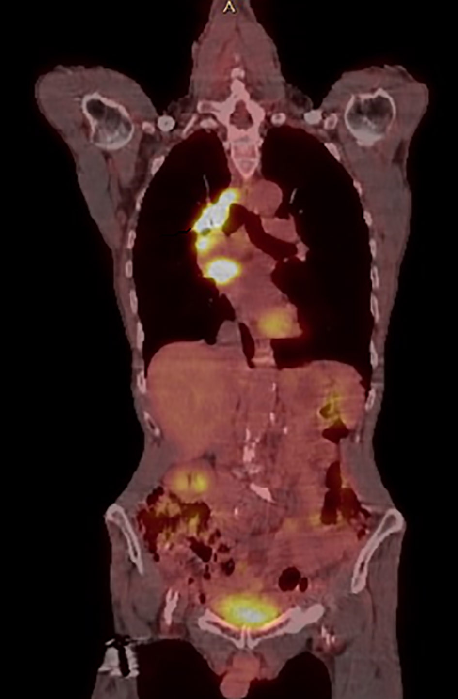
Figure 1. PET/CT longitudinal view showing abnormal uptake throughout right mediastinum and LA. PET/CT: positron emission tomography/computerized tomography; LA: left atrium.
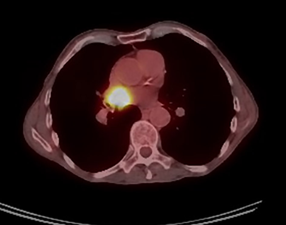
Figure 2. PET/CT cross-sectional view showing abnormal uptake throughout the right mediastinum and the LA. PET/CT: positron emission tomography/computerized tomography; LA: left atrium.
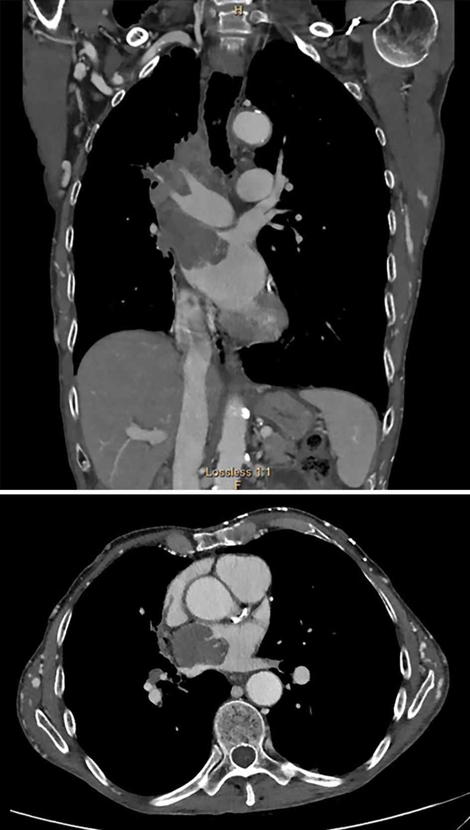
Figure 3. Right hilar mass eroding into the LA. Notice multiple chest wall collaterals secondary to SVC occlusion. LA: left atrium; SVC: superior vena cava.
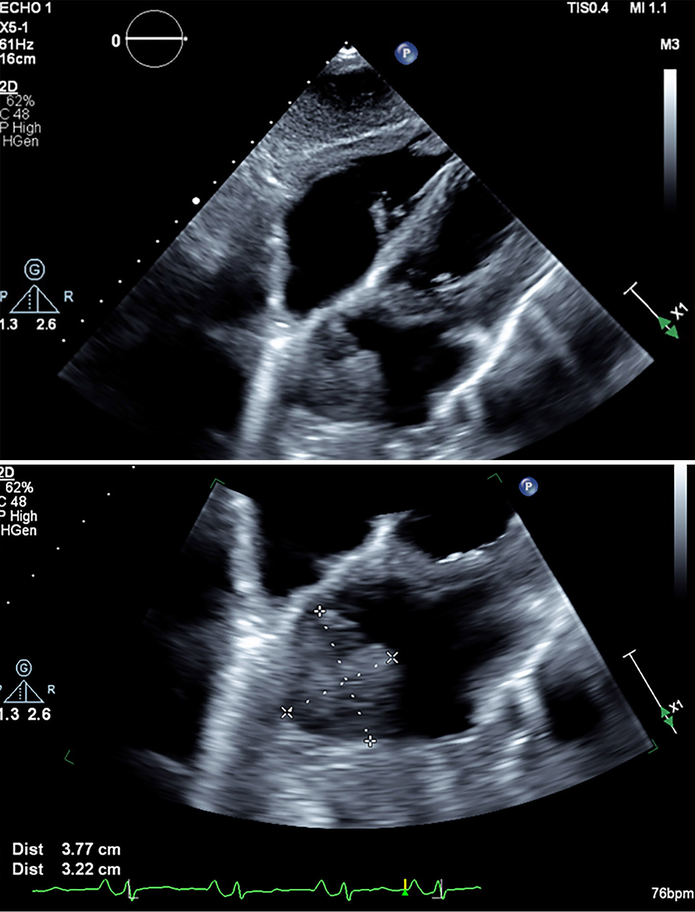
Figure 4. Subcostal views showing large left atrial mass measured 3.8 × 3.2 cm in left atrial cavity.
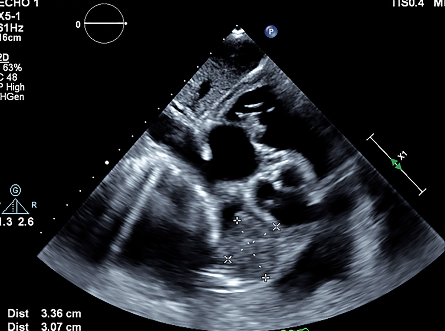
Figure 5. Subcostal five-chamber view showing almost complete obliteration of left atrial cavity.
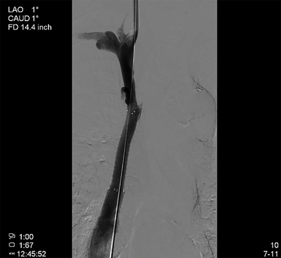
Figure 6. Angiographic image of SVC after stenting with 10 × 60 mm Fluency stent. SVC: superior vena cava.
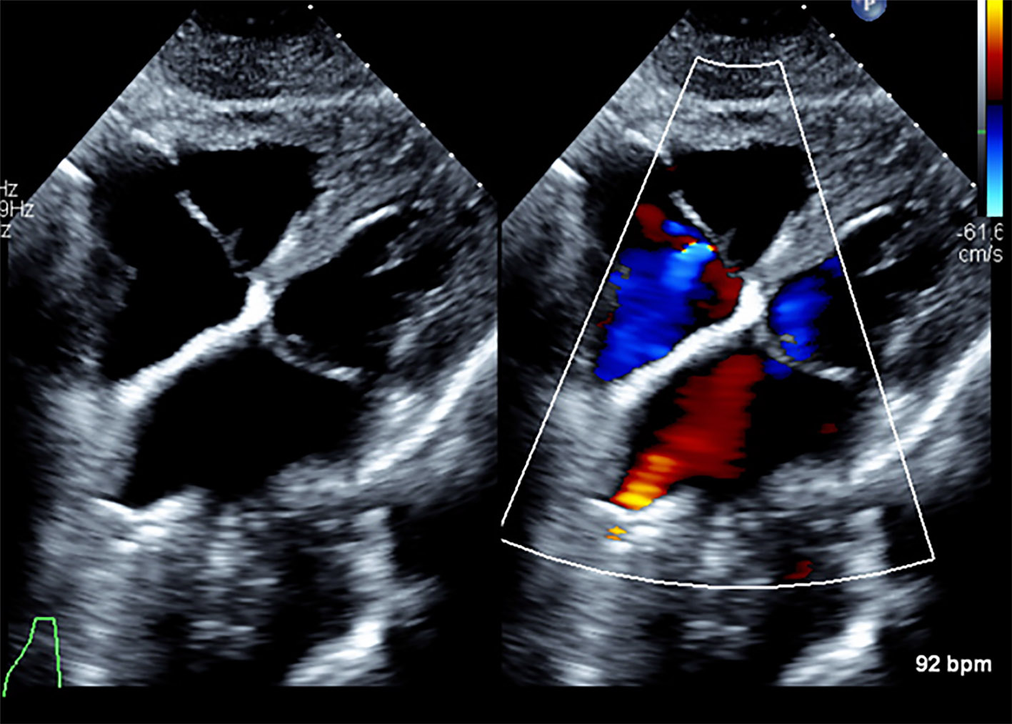
Figure 7. Resolution of left atrial tumor thrombus on echo 6 weeks after chemo- and radiotherapy.
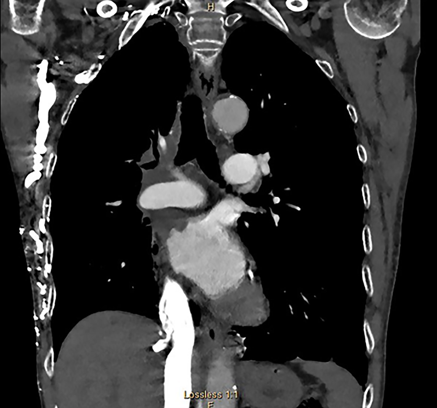
Figure 8. Decrease in size of right mediastinal and left atrial mass. Notice multiple chest wall collaterals secondary to SVC occlusion. SVC: superior vena cava.