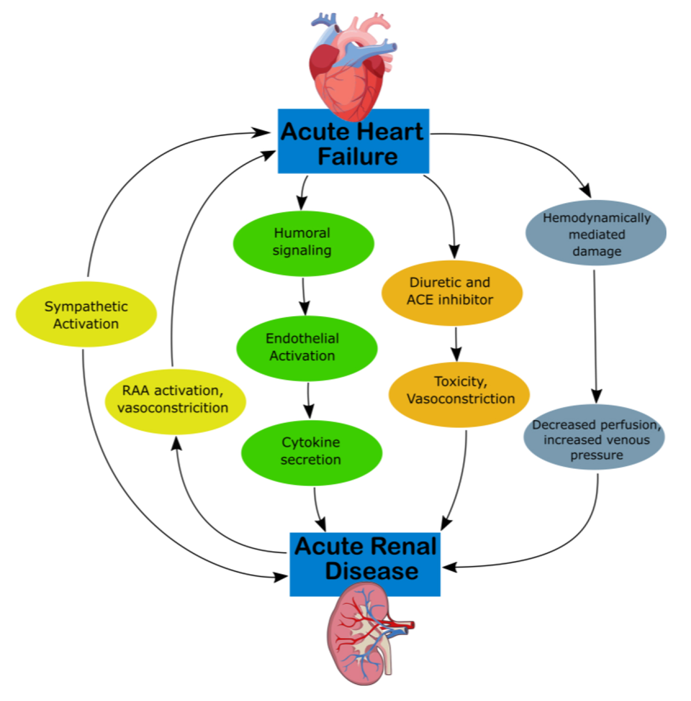
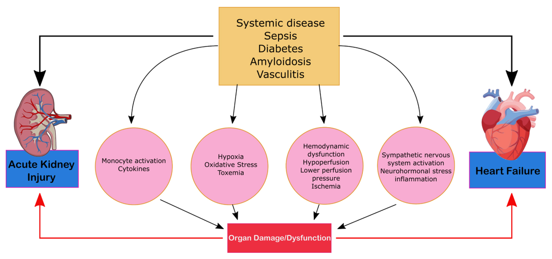
Figure 1. Mechanism of damage in CRS-1. ACE, angiotensin-converting enzyme; CKD, chronic kidney disease; CRS, cardiorenal syndrome; RAA, rennin-angiotensin-aldosterone.
| Cardiology Research, ISSN 1923-2829 print, 1923-2837 online, Open Access |
| Article copyright, the authors; Journal compilation copyright, Cardiol Res and Elmer Press Inc |
| Journal website http://www.cardiologyres.org |
Review
Volume 11, Number 2, April 2020, pages 76-88
An Update on the Pathophysiology and Treatment of Cardiorenal Syndrome
Figures
Tables
| Cardiorenal types | Characteristics | Causes of morbidity |
|---|---|---|
| Type 1 (acute cardiorenal) | Acute cardiac impairment leading to acute kidney injury (AKI) | Cardiogenic shock and AKI, acute decompensated heart failure (ADHF) resulting in AKI |
| Type 2 (chronic cardiorenal) | Chronic cardiac impairment leading to renal impairment | Chronic heart failure |
| Type 3 (acute renocardiac) | AKI leading to cardiac impairment | Heart failure in the setting of AKI from volume overload, inflammatory surge and accompanying metabolic disturbances |
| Type 4 (chronic renocardiac) | Chronic kidney disease (CKD) leading to cardiac impairment | Myocardial remodeling and heart failure from CKD-associated cardiomyopathy |
| Type 5 (secondary cardiorenal) | Systemic condition leading to both cardiac and renal impairment | Diabetes, amyloidosis and sepsis |
| Biomarkers | Characteristics of presentation/site of origin |
|---|---|
| Molecular biomarkers | |
| Cardiac troponin I (cTnI) | Myocardial injury |
| B-type natriuretic peptide (BNP) | Myocardial stretching |
| sST2 | Member of interleukin (IL)-1 family of receptors |
| Indoxyl sulfate | Extracellular signal-regulated kinase (ERK), p38 mitogen-activated protein kinase (MAPK) and nuclear factor-kappa B (NF-κB) |
| N-terminal propeptide of type III collagen (PIIINP) | Connective tissue injury |
| Physiological biomarkers | |
| Echocardiogram | Abnormal left ventricular hypertrophy |
| Central venous pressure | |
| Pericardial effusion | |
| Valvular stenosis | |
| Myocardial injury | |
| Fluid overfill | |
| Valvular calcification | |
| Doppler | Intraparenchymal blood flow that is associated |
| Ultrasound | Fluid overload |
| Chest radiograph | Cardiomegaly |
| Interstitial edema | |
| Enlarged pulmonary artery | |
| Pleural effusion | |
| Prominent superior vena cava | |
| Kerley line | |
| Kidney biomarkers | |
| Serum creatinine | Skeletal muscle |
| Albuminuria | Marker of glomerular integrity/procalcitonin (PCT) disruption |
| Kidney injury molecule (KIM-1) | Type 1 cell membrane glycoprotein expressed in regenerating PCT epithelium |
| Liver type fatty acid binding protein | Tubular injury |
| IL-18 | Cytokine mediating inflammation and AKI through the NF-κB pathway |
| Advanced glycation end products (AGE) | Improper renal clearance, myoc |
| Physiological biomarkers | |
| Ultrasound | Kidney enlargement |
| Thin and hyperechogenic cortex | |
| Small dilation of the urinary tract | |
| Parapelvic and subcortical cysts | |
| Doppler | Intraparenchymal blood flow |